- Published on
Dermatology - Vesicles and Bullae
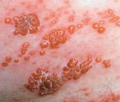
Vesicle is derived from the Latin word "vesicula," which means "little bladder," while bulla is derived from the Latin word "bulla," which means "bubble."
A skin eruption characterized by the presence of small fluid-filled sacs is referred to as a vesicular exanthem, whereas a skin eruption characterized by the presence of large fluid-filled sacs is referred to as a bullous exanthem.
A vesicle is a small cavity, measuring less than 0.5 cm, that is raised and contains fluid. On the other hand, a bulla is a larger cavity, measuring more than 0.5 cm, that is also raised and contains fluid.
One can feel vesicles and bulla upon palpation.
Vesicles exhibit a dome-shaped structure and can either be umbilicated (similar to herpes simplex) or flaccid.
Frequently, the roof of a vesicle or bulla is sufficiently thin to be translucent.
The contents of the vesicle/bulla are frequently visible. The vesicles containing serum exhibit a yellowish hue, whereas those containing blood range in color from red to black. Vesicles and bullae form as a result of a separation in different layers of the outermost layer of the skin. This separation might occur just below the top layer, inside the visible layer of the skin, or at the boundary between the outermost layer and the layer beneath it. As vesicles/bullae are consistently located near the surface, they possess a clear and distinct appearance.
Vesicle is derived from the Latin word "vesicula," which means "little bladder," while bulla is derived from the Latin word "bulla," which means "bubble."
A skin eruption characterized by the presence of small fluid-filled sacs is referred to as a vesicular exanthem, whereas a skin eruption characterized by the presence of large fluid-filled sacs is referred to as a bullous exanthem.
A vesicle is a small cavity, measuring less than 0.5 cm, that is raised and contains fluid. On the other hand, a bulla is a larger cavity, measuring more than 0.5 cm, that is also raised and contains fluid.
One can feel vesicles and bulla upon palpation.
Vesicles exhibit a dome-shaped structure and can either be umbilicated (similar to herpes simplex) or flaccid.
Frequently, the roof of a vesicle or bulla is sufficiently thin to be translucent.
The contents of the vesicle/bulla are frequently visible. The vesicles containing serum exhibit a yellowish hue, whereas those containing blood range in color from red to black. Vesicles and bullae form as a result of a separation in different layers of the outermost layer of the skin. This separation might occur just below the top layer, inside the visible layer of the skin, or at the boundary between the outermost layer and the layer beneath it. As vesicles/bullae are consistently located near the surface, they possess a clear and distinct appearance.
- Published on
Dermatology - Crust
The term "crust" originates from the Latin word for rind, bark, or shell.
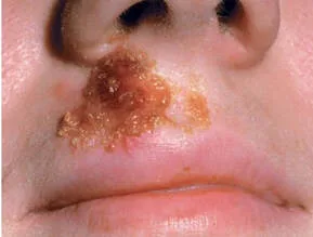
Crusts form when serum, blood, or purulent discharge dries on the surface of the skin.
• Crusts can vary in thickness and texture, ranging from thin, delicate, and crumbly to thick and firmly attached. Crusts can be felt by touch.
Crusts appear yellow when they are created from dried serum, green or yellow green when they are formed from purulent exudate, and brown, dark red, or black when they are formed from blood. Superficial crusts manifest as honey-colored, fragile, shiny particles on the surface. If the exudate covers the entire epidermis, the crusts can become thick and stick to the skin. If this is accompanied by tissue death in the deeper layers (such as the dermis), the disease is referred to as ecthyma.
The term "crust" originates from the Latin word for rind, bark, or shell.
Crusts form when serum, blood, or purulent discharge dries on the surface of the skin.
• Crusts can vary in thickness and texture, ranging from thin, delicate, and crumbly to thick and firmly attached. Crusts can be felt by touch.
Crusts appear yellow when they are created from dried serum, green or yellow green when they are formed from purulent exudate, and brown, dark red, or black when they are formed from blood. Superficial crusts manifest as honey-colored, fragile, shiny particles on the surface. If the exudate covers the entire epidermis, the crusts can become thick and stick to the skin. If this is accompanied by tissue death in the deeper layers (such as the dermis), the disease is referred to as ecthyma.
- Published on
Dermatology - Scales
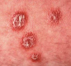
The term "squames" is derived from the Latin word meaning "scale." A skin condition characterized by the presence of raised lesions with scales is referred to as a papulosquamous exanthem. Scales are thin layers of the outermost layer of the skin, known as the stratum corneum.
They can vary in size, ranging from huge structures like membranes to minute particles like dust. They can also be described as pityriasiform, which means resembling bran, and can either be adhering or loose.
Scales can manifest either independently or superimposed on other types of lesions.
Palpation of scales is optional.
Scales can have either well-defined or indistinct boundaries.
The term "squames" is derived from the Latin word meaning "scale." A skin condition characterized by the presence of raised lesions with scales is referred to as a papulosquamous exanthem. Scales are thin layers of the outermost layer of the skin, known as the stratum corneum.
They can vary in size, ranging from huge structures like membranes to minute particles like dust. They can also be described as pityriasiform, which means resembling bran, and can either be adhering or loose.
Scales can manifest either independently or superimposed on other types of lesions.
Palpation of scales is optional.
Scales can have either well-defined or indistinct boundaries.
- Published on
Dermatology - Plaque
The word "plaque" is the French equivalent of the English word "plate."
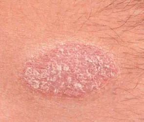
Plaques are created when papules merge together to produce bigger, typically flat-topped, well-defined elevations on the skin surface. Plaques cover a relatively considerable area of the skin compared to their height. Plaques can be felt when touched.
Plaques typically exhibit clear boundaries. Lichenification refers to the presence of a big, poorly defined patch of thicker skin with exaggerated skin marks. A patch is a slightly raised plaque, which is a type of skin lesion that falls between a macule (flat, discolored spot) and a plaque (larger, raised area).
The word "plaque" is the French equivalent of the English word "plate."
Plaques are created when papules merge together to produce bigger, typically flat-topped, well-defined elevations on the skin surface. Plaques cover a relatively considerable area of the skin compared to their height. Plaques can be felt when touched.
Plaques typically exhibit clear boundaries. Lichenification refers to the presence of a big, poorly defined patch of thicker skin with exaggerated skin marks. A patch is a slightly raised plaque, which is a type of skin lesion that falls between a macule (flat, discolored spot) and a plaque (larger, raised area).
- Published on
Dermatology - Nodule
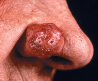
The term "nodule" is derived from the Latin word "nodulus," which means a little knot. A nodule is a firm, circular, or oval-shaped abnormality that is bigger than a papule. Palpable nodules may be present, affecting the epidermis, dermis, or subcutaneous tissue.
Nodules can have either a clear boundary (superficial) or an unclear boundary (deep). When they are located in the layer of tissue just below the skin, they are generally more easily detected by touch than by sight. Nodules may exhibit either firm or pliable characteristics when examined by touch.
They can have a domed and smooth morphology or possess a surface characterized by warts or a central depression resembling a crater.
The distinction between a nodule and a papule lies on their respective levels of involvement and size. Nodules arise due to the presence of inflammatory infiltrates, neoplasms, or metabolic deposits in the dermis or subcutaneous tissue.
The term "nodule" is derived from the Latin word "nodulus," which means a little knot. A nodule is a firm, circular, or oval-shaped abnormality that is bigger than a papule. Palpable nodules may be present, affecting the epidermis, dermis, or subcutaneous tissue.
Nodules can have either a clear boundary (superficial) or an unclear boundary (deep). When they are located in the layer of tissue just below the skin, they are generally more easily detected by touch than by sight. Nodules may exhibit either firm or pliable characteristics when examined by touch.
They can have a domed and smooth morphology or possess a surface characterized by warts or a central depression resembling a crater.
The distinction between a nodule and a papule lies on their respective levels of involvement and size. Nodules arise due to the presence of inflammatory infiltrates, neoplasms, or metabolic deposits in the dermis or subcutaneous tissue.
- Published on
Infectious Diseases and Microbiology - Taeniasis (Beef Tapeworm )
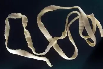
Beef tapeworm infection, or taeniasis
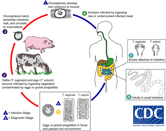
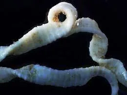
Beef tapeworms, or Cestodes, are known by the name Taenia saginata.
One or more hermaphrodite proglottids can be found in Strobila.
Cattle are the intermediate host; humans are the final host. When undercooked beef is consumed, infections spread to humans. T saginata does not cause cysticercosis.
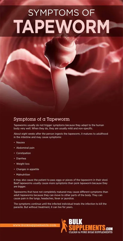
Adult tapeworm infections are asymptomatic or present with minor gastrointestinal symptoms.
The scolex is how adult tapeworms stay connected to the intestine, and they can reach lengths of up to 10 meters.
It is diagnostic to see proglottids with 15–30 uterine branches or eggs in the perianal area or feces.
Praziquantel is frequently used as a medication. To prevent infection in animals, prevent undercooked beef and dispose of human waste properly.
Beef tapeworm infection, or taeniasis
Beef tapeworms, or Cestodes, are known by the name Taenia saginata.
One or more hermaphrodite proglottids can be found in Strobila.
Cattle are the intermediate host; humans are the final host. When undercooked beef is consumed, infections spread to humans. T saginata does not cause cysticercosis.
Adult tapeworm infections are asymptomatic or present with minor gastrointestinal symptoms.
The scolex is how adult tapeworms stay connected to the intestine, and they can reach lengths of up to 10 meters.
It is diagnostic to see proglottids with 15–30 uterine branches or eggs in the perianal area or feces.
Praziquantel is frequently used as a medication. To prevent infection in animals, prevent undercooked beef and dispose of human waste properly.
- Published on
Infectious Diseases and Microbiology - Diphyllobothriasis ( Diphyllobothrium latum)
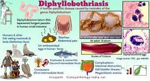
Diphyllobothrium latum is a type of cestode, which is a segmented flatworm. It is commonly known as the broad fish tapeworm.
The strobila consists of one or many hermaphroditic proglottids.
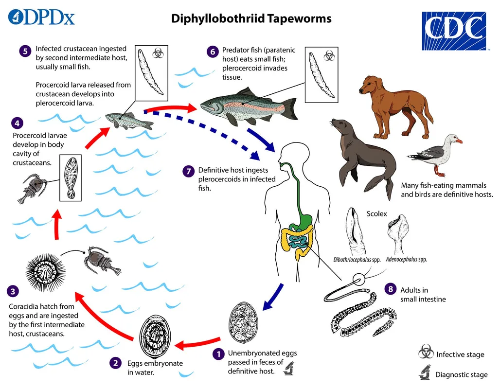
Humans are the primary host. D latum utilizes two consecutive intermediate hosts: a freshwater copepod known as Cyclops and a freshwater fish. Infection occurs as a consequence of consuming freshwater fish that is undercooked, pickled, or uncooked.
The majority of infections do not show any symptoms; nevertheless, when there is a scarcity of vitamin B12, it can cause a shortage in B12, leading to megaloblastic anemia and a range of neurological symptoms.
Adult tapeworms maintain their attachment to the intestine through the scolex and can reach a length of up to 13 meters.
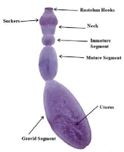
The eggs of D latum have an elongated shape and possess a lidlike operculum. The proglottids exhibit a greater width compared to their length, featuring a uterine branch pattern arranged in a rosette formation.
Praziquantel is administered for therapeutic purposes. Prevention include refraining from consuming undercooked fish and implementing precautions to avoid contamination of freshwater lakes by human feces.
Diphyllobothrium latum is a type of cestode, which is a segmented flatworm. It is commonly known as the broad fish tapeworm.
The strobila consists of one or many hermaphroditic proglottids.
Humans are the primary host. D latum utilizes two consecutive intermediate hosts: a freshwater copepod known as Cyclops and a freshwater fish. Infection occurs as a consequence of consuming freshwater fish that is undercooked, pickled, or uncooked.
The majority of infections do not show any symptoms; nevertheless, when there is a scarcity of vitamin B12, it can cause a shortage in B12, leading to megaloblastic anemia and a range of neurological symptoms.
Adult tapeworms maintain their attachment to the intestine through the scolex and can reach a length of up to 13 meters.
The eggs of D latum have an elongated shape and possess a lidlike operculum. The proglottids exhibit a greater width compared to their length, featuring a uterine branch pattern arranged in a rosette formation.
Praziquantel is administered for therapeutic purposes. Prevention include refraining from consuming undercooked fish and implementing precautions to avoid contamination of freshwater lakes by human feces.
- Published on
Infectious Diseases and Microbiology - Echinococcosis / Hydatid Cyst Disease ( Echinococcus granulosus)
Hydatid cyst disease, also known as echinococcosis
A cestode, or segmented flatworm, is Echinococcus granulosus.
A scolex and three proglottids make it extremely diminutive.
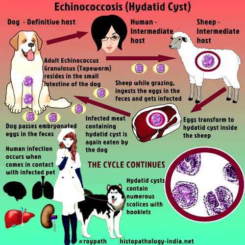
Dogs are the last hosts, while sheep play a more intermediary role. The viscera of sick sheep can infect dogs. When people swallow eggs that have been contaminated with dog excrement, they can contract an infection.
Cysts can form in a variety of organs, and humans are unwitting intermediary hosts. People cannot grow adult worms.
Hidatid cyst disease is caused by E granulosus. Based on where it grows, this can not cause any symptoms at all or show a wide range of symptoms in the brain, liver, or lungs.
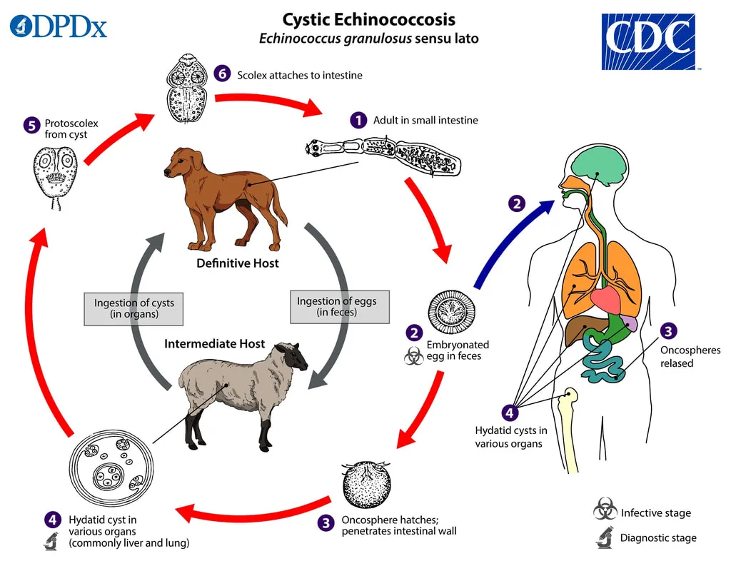
Eggs hatch into embryos, which then invade cells in the intestines and make their way into the circulation. During their development, larvae build a cyst (a hydatid cyst) that is filled with fluid and contains protoscoleces in brood capsules. When discovered in different organs, the hydatid cysts—which can be as big as 20 cm in diameter—cause the observed pathology. When the cyst bursts, it releases infectious protoscoleces into the bloodstream, which can trigger anaphylaxis.
A diagnosis of hydatid cysts can be made using serologic testing or imaging techniques like X-ray or CT.
A course of albendazole and meticulous surgical excision of the hydatid cyst constitute the treatment.Hygienic methods to avoid dog excrement contamination of food and water, deworming of dogs, and not feeding dogs sheep viscera are all ways to prevent this disease.
Hydatid cyst disease, also known as echinococcosis
A cestode, or segmented flatworm, is Echinococcus granulosus.
A scolex and three proglottids make it extremely diminutive.
Dogs are the last hosts, while sheep play a more intermediary role. The viscera of sick sheep can infect dogs. When people swallow eggs that have been contaminated with dog excrement, they can contract an infection.
Cysts can form in a variety of organs, and humans are unwitting intermediary hosts. People cannot grow adult worms.
Hidatid cyst disease is caused by E granulosus. Based on where it grows, this can not cause any symptoms at all or show a wide range of symptoms in the brain, liver, or lungs.
Eggs hatch into embryos, which then invade cells in the intestines and make their way into the circulation. During their development, larvae build a cyst (a hydatid cyst) that is filled with fluid and contains protoscoleces in brood capsules. When discovered in different organs, the hydatid cysts—which can be as big as 20 cm in diameter—cause the observed pathology. When the cyst bursts, it releases infectious protoscoleces into the bloodstream, which can trigger anaphylaxis.
A diagnosis of hydatid cysts can be made using serologic testing or imaging techniques like X-ray or CT.
A course of albendazole and meticulous surgical excision of the hydatid cyst constitute the treatment.Hygienic methods to avoid dog excrement contamination of food and water, deworming of dogs, and not feeding dogs sheep viscera are all ways to prevent this disease.
- Published on
Infectious Diseases and Microbiology - Cestode induced Diarrhea ( Hymenolepis nana)
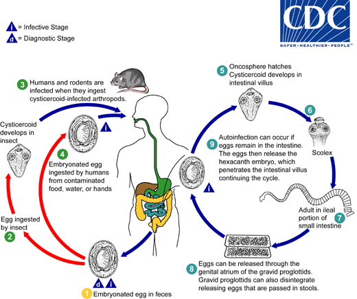
Cestode-induced Diarrhea is caused by Hymenolepis nana, a type of segmented flatworm known as a dwarf tapeworm. This tapeworm is the tiniest among adult tapeworms. The complete life cycle occurs within people.
The etiology of this condition involves humans as the definitive host, with no necessary intermediate hosts. The mode of transmission is through the ingestion of fecal matter.
Particularly common among children. Hymenolepis nana is a prevalent tapeworm infection, particularly in the southeastern region of the United States.
Infections typically do not show symptoms when there are just a few worms present, but as the number of worms increases, gastrointestinal symptoms become more apparent.
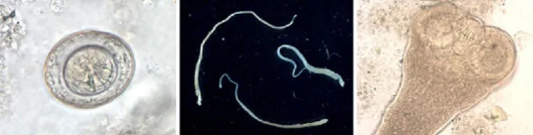
Eggs are highly contagious. After being consumed, the larvae undergo development and attach themselves to the intestine. The adult worms are of diminutive size, measuring 2–5 cm in length, and generate eggs that are excreted in feces. The host can experience autoinfection, leading to a significant increase in the number of worms, often reaching several hundred.
Detection of H nana eggs (including a six-hooked embryo) in stool is indicative of the presence of the parasite.
The standard course of treatment usually consists of praziquantel. The most effective approach to prevention is implementing measures that eradicate the transmission of fecal matter through the mouth.
Cestode-induced Diarrhea is caused by Hymenolepis nana, a type of segmented flatworm known as a dwarf tapeworm. This tapeworm is the tiniest among adult tapeworms. The complete life cycle occurs within people.
The etiology of this condition involves humans as the definitive host, with no necessary intermediate hosts. The mode of transmission is through the ingestion of fecal matter.
Particularly common among children. Hymenolepis nana is a prevalent tapeworm infection, particularly in the southeastern region of the United States.
Infections typically do not show symptoms when there are just a few worms present, but as the number of worms increases, gastrointestinal symptoms become more apparent.
Eggs are highly contagious. After being consumed, the larvae undergo development and attach themselves to the intestine. The adult worms are of diminutive size, measuring 2–5 cm in length, and generate eggs that are excreted in feces. The host can experience autoinfection, leading to a significant increase in the number of worms, often reaching several hundred.
Detection of H nana eggs (including a six-hooked embryo) in stool is indicative of the presence of the parasite.
The standard course of treatment usually consists of praziquantel. The most effective approach to prevention is implementing measures that eradicate the transmission of fecal matter through the mouth.
- Published on
Infectious Diseases and Microbiology - Schistosomiasis
( Schistosoma mansoni)
Schistosomiasis is a parasitic disease.
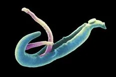
Schistosoma mansoni is a nonsegmented flatworm or fluke belonging to the Trematode class which causes schistosomiasis.
There are three prevalent species of human schistosomes: S mansoni, S japonicum, and S haematobium.
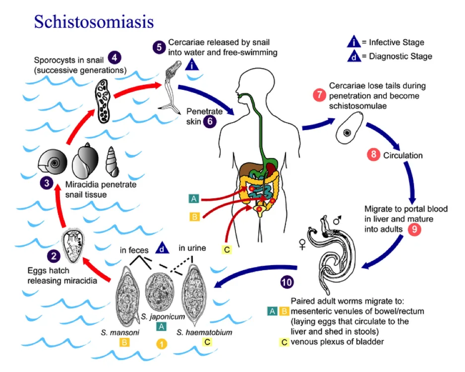
Snails serve as intermediary hosts. Humans are the ultimate hosts. Cercariae enter the human skin, mature into adults, and deposit eggs that are excreted in urine (haematobium) or stool (mansoni and japonicum).
The regions where the disease is commonly found are Africa, South America, the Middle East, the West Indies, and Puerto Rico for S. mansoni; China, Japan, and the Philippines for S. japonicum; and Africa, the Middle East, and India for S. haematobium. Swimmer's itch occurs when cercariae from avian and mammalian species of Schistosoma penetrate the skin.
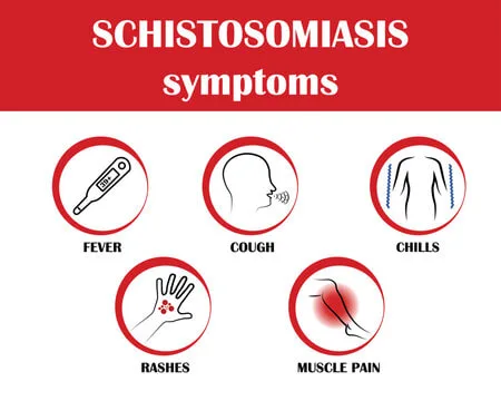
Following the entrance of cercariae into the skin, a temporary rash characterized by itching and the presence of little raised bumps may develop. Acute symptoms consist of elevated body temperature and shivering. The prevalence of chronic diseases in adults varies depending on their geographical region. Schistosoma mansoni and Schistosoma japonicum result in abdominal pain, bloody diarrhea, and hepatosplenomegaly. Schistosoma haematobium induces the symptoms of blood in urine, painful urination, and blockage, and is linked to bladder cancer.
Following the invasion of the skin, the organism infiltrates the bloodstream and then travels to the lungs. Each of the three primary human schistosome parasites resides within a specific region of the venous plexus that drains either the intestines or bladder, depending on the particular species. Specifically, S mansoni inhabits the mesenteric veins of the colon, S japonicum resides in the mesenteric veins of the small bowel and colon, and S haematobium is found in the veins surrounding the bladder and pelvic organs. Additionally, all three parasites are capable of producing eggs.
The pathogenesis of this condition arises from the immune system's reaction to the eggs, leading to the formation of abscesses, fibrosis, granulomas, and scarring. The eggs secrete hydrolytic enzymes that degrade tissue and facilitate the shedding process.
The presence of eggs in pee or stool is a distinctive trait.
Praziquantel is an effective therapy option. Prevention entails implementing effective waste management practices, refraining from swimming in freshwater bodies located in regions with a high prevalence of the disease, and eliminating snail populations.
( Schistosoma mansoni)
Schistosomiasis is a parasitic disease.
Schistosoma mansoni is a nonsegmented flatworm or fluke belonging to the Trematode class which causes schistosomiasis.
There are three prevalent species of human schistosomes: S mansoni, S japonicum, and S haematobium.
Snails serve as intermediary hosts. Humans are the ultimate hosts. Cercariae enter the human skin, mature into adults, and deposit eggs that are excreted in urine (haematobium) or stool (mansoni and japonicum).
The regions where the disease is commonly found are Africa, South America, the Middle East, the West Indies, and Puerto Rico for S. mansoni; China, Japan, and the Philippines for S. japonicum; and Africa, the Middle East, and India for S. haematobium. Swimmer's itch occurs when cercariae from avian and mammalian species of Schistosoma penetrate the skin.
Following the entrance of cercariae into the skin, a temporary rash characterized by itching and the presence of little raised bumps may develop. Acute symptoms consist of elevated body temperature and shivering. The prevalence of chronic diseases in adults varies depending on their geographical region. Schistosoma mansoni and Schistosoma japonicum result in abdominal pain, bloody diarrhea, and hepatosplenomegaly. Schistosoma haematobium induces the symptoms of blood in urine, painful urination, and blockage, and is linked to bladder cancer.
Following the invasion of the skin, the organism infiltrates the bloodstream and then travels to the lungs. Each of the three primary human schistosome parasites resides within a specific region of the venous plexus that drains either the intestines or bladder, depending on the particular species. Specifically, S mansoni inhabits the mesenteric veins of the colon, S japonicum resides in the mesenteric veins of the small bowel and colon, and S haematobium is found in the veins surrounding the bladder and pelvic organs. Additionally, all three parasites are capable of producing eggs.
The pathogenesis of this condition arises from the immune system's reaction to the eggs, leading to the formation of abscesses, fibrosis, granulomas, and scarring. The eggs secrete hydrolytic enzymes that degrade tissue and facilitate the shedding process.
The presence of eggs in pee or stool is a distinctive trait.
Praziquantel is an effective therapy option. Prevention entails implementing effective waste management practices, refraining from swimming in freshwater bodies located in regions with a high prevalence of the disease, and eliminating snail populations.