- Published on
Dermatology - Macule
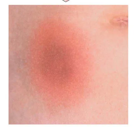
The term "macule" is derived from the Latin word for "spot." A skin eruption characterized by flat, discolored patches is referred to as a macular exanthem.
A macule is a defined region on the skin that exhibits a shift in color without any raised or sunken areas.Macules are characterized by their flat nature and lack of palpability.
Macules might have distinct boundaries (sharply defined) or spread out. • Macules can vary in size or color. Macules can have pigmentation in shades of brown, black, or blue, or they might be skin-colored (amelanotic) or red.
Diagnosis
Diascopy, which involves applying pressure from a glass slide on the edge of a red macule, allows for the detection of red blood cells. If the redness persists when pressure is applied to the slide, the lesion is classified as purpuric, indicating that it is caused by the leakage of red blood cells. Conversely, if the redness vanishes, the lesion is attributed to vascular dilation.
The term "macule" is derived from the Latin word for "spot." A skin eruption characterized by flat, discolored patches is referred to as a macular exanthem.
A macule is a defined region on the skin that exhibits a shift in color without any raised or sunken areas.Macules are characterized by their flat nature and lack of palpability.
Macules might have distinct boundaries (sharply defined) or spread out. • Macules can vary in size or color. Macules can have pigmentation in shades of brown, black, or blue, or they might be skin-colored (amelanotic) or red.
Diagnosis
Diascopy, which involves applying pressure from a glass slide on the edge of a red macule, allows for the detection of red blood cells. If the redness persists when pressure is applied to the slide, the lesion is classified as purpuric, indicating that it is caused by the leakage of red blood cells. Conversely, if the redness vanishes, the lesion is attributed to vascular dilation.
- Published on
Dermatology - Papule
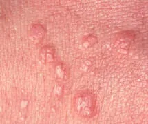
A papule is a medical term derived from Latin that refers to a small, raised skin lesion, commonly known as a pimple. A skin eruption characterized by little raised lesions is referred to as a papular exanthem.
Papular exanthems can be classified as either "lichenoid" when they are gathered together, or "dispersed" when they are spread apart.
A papule is a small, raised, firm skin lesion, often measuring less than 0.5 cm in diameter. The majority of the lesion is raised above the level of the surrounding skin, rather than being located deep within it.
• Papules are rounded, cone-shaped, or raised with a flat surface that can be felt. • The elevation is caused by the accumulation of substances within the lesion, such as metabolic or locally generated deposits, localized cellular infiltrates, or inflammatory cellular components.
Papules can exhibit either well-defined (sharply marginated) or ill-defined (diffuse) characteristics. Superficial papules typically have clear boundaries, while deeper dermal papules display less apparent edges.
A papule is a medical term derived from Latin that refers to a small, raised skin lesion, commonly known as a pimple. A skin eruption characterized by little raised lesions is referred to as a papular exanthem.
Papular exanthems can be classified as either "lichenoid" when they are gathered together, or "dispersed" when they are spread apart.
A papule is a small, raised, firm skin lesion, often measuring less than 0.5 cm in diameter. The majority of the lesion is raised above the level of the surrounding skin, rather than being located deep within it.
• Papules are rounded, cone-shaped, or raised with a flat surface that can be felt. • The elevation is caused by the accumulation of substances within the lesion, such as metabolic or locally generated deposits, localized cellular infiltrates, or inflammatory cellular components.
Papules can exhibit either well-defined (sharply marginated) or ill-defined (diffuse) characteristics. Superficial papules typically have clear boundaries, while deeper dermal papules display less apparent edges.
- Published on
Infectious Diseases and Microbiology - Toxocariasis / Visceral Larva Migrans (Toxocara canis and Toxocara cati )
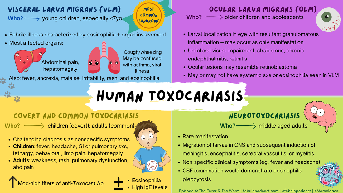
Toxocariasis, also known as Visceral Larva Migrans
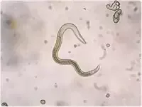
Toxocara canis and Toxocara cati are both species of Nematode, which are a type of roundworm.
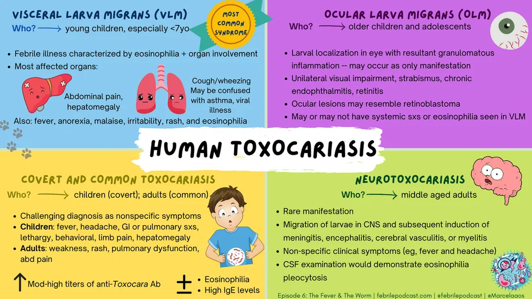
The tissue manifestations are referred to as Visceral larva migrans.
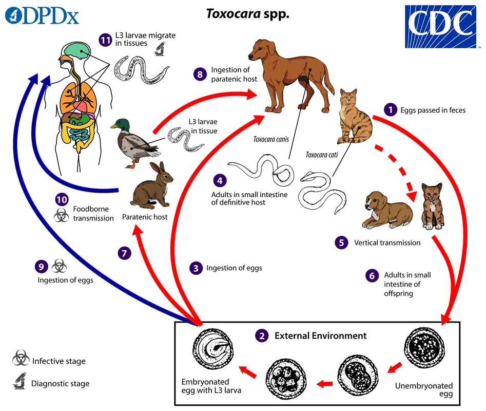
Humans are terminal hosts. Dogs (T canis) and cats (T cati) serve as decisive hosts. Dogs and cats excrete Toxocara eggs in their feces. Humans, particularly youngsters aged 1-4 with a background of pica, acquire the infection by consuming eggs.
A significant number of persons are without symptoms. The symptoms of larval infestation might vary depending on their location. Some individuals may have gastrointestinal pain, rash, fever, enlarged liver, retinal involvement resulting in blindness, or manifestations affecting the heart, respiratory system, or central nervous system.
Upon ingestion, eggs give rise to larvae that then move to various organs, such as the lungs, liver, kidney, heart, eyes, muscle, and central nervous system (CNS). The larvae undergo encystation, transforming into second-stage larvae. The larvae migration results in the formation of tracts characterized by hemorrhagic necrosis, as well as infiltration of eosinophils and lymphocytes. Granulomas can form around deteriorating larvae. The development of the disease is caused by the intensity of the inflammation and the specific site and quantity of the deceased larvae.
Elevated levels of hypereosinophilia and hypergammaglobulinemia, along with increased titers of isohemagglutinin against the A and B blood group antigens, are indicative of a probable infection. The diagnostic process entails serologic testing, analysis of tissue biopsies for larvae, and evaluation of clinical signs and symptoms. Stool samples can be analyzed to detect eggs in pets.
Mebendazole and albendazole are utilized for therapeutic purposes. Prevention entails the practice of deworming pets and maintaining proper hygiene for pets.
Toxocariasis, also known as Visceral Larva Migrans
Toxocara canis and Toxocara cati are both species of Nematode, which are a type of roundworm.
The tissue manifestations are referred to as Visceral larva migrans.
Humans are terminal hosts. Dogs (T canis) and cats (T cati) serve as decisive hosts. Dogs and cats excrete Toxocara eggs in their feces. Humans, particularly youngsters aged 1-4 with a background of pica, acquire the infection by consuming eggs.
A significant number of persons are without symptoms. The symptoms of larval infestation might vary depending on their location. Some individuals may have gastrointestinal pain, rash, fever, enlarged liver, retinal involvement resulting in blindness, or manifestations affecting the heart, respiratory system, or central nervous system.
Upon ingestion, eggs give rise to larvae that then move to various organs, such as the lungs, liver, kidney, heart, eyes, muscle, and central nervous system (CNS). The larvae undergo encystation, transforming into second-stage larvae. The larvae migration results in the formation of tracts characterized by hemorrhagic necrosis, as well as infiltration of eosinophils and lymphocytes. Granulomas can form around deteriorating larvae. The development of the disease is caused by the intensity of the inflammation and the specific site and quantity of the deceased larvae.
Elevated levels of hypereosinophilia and hypergammaglobulinemia, along with increased titers of isohemagglutinin against the A and B blood group antigens, are indicative of a probable infection. The diagnostic process entails serologic testing, analysis of tissue biopsies for larvae, and evaluation of clinical signs and symptoms. Stool samples can be analyzed to detect eggs in pets.
Mebendazole and albendazole are utilized for therapeutic purposes. Prevention entails the practice of deworming pets and maintaining proper hygiene for pets.
- Published on
Infectious Diseases and Microbiology - Cysticercosis with Neurological Involvement (Taenia solium )
Infectious Diseases and Microbiology - Cysticercosis with Neurological Involvement (Taenia solium )
Neurological involvement in cysticercosis
Taenia solium is a type of segmented flatworm known as a Cestode. It is commonly referred to as a pork tapeworm.
The strobila consists of one or many hermaphroditic proglottids.
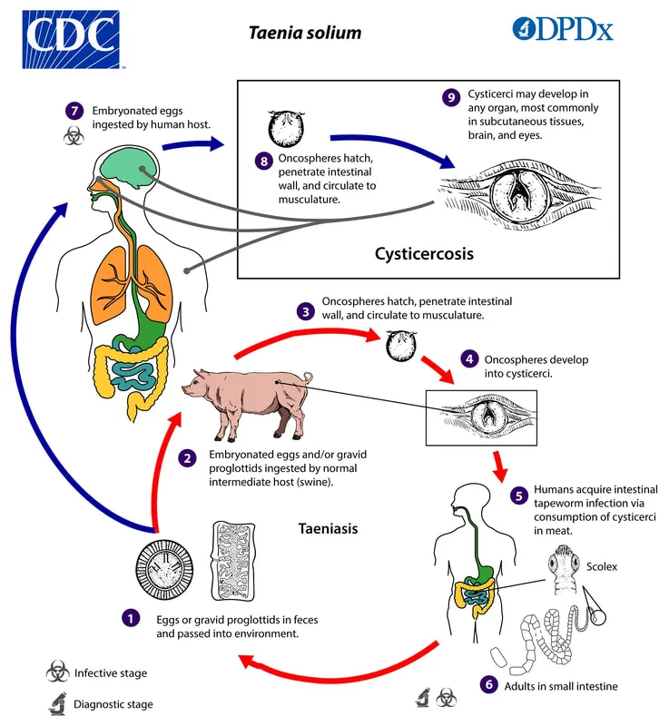
Humans serve as the primary host, while pigs act as the intermediary host. Pigs consume eggs, and the embryo penetrates the intestinal wall, enters the bloodstream, and disseminates to skeletal muscle. Infection in humans occurs when they consume undercooked pork that is contaminated with encysted larvae. Tapeworms mature within the gastrointestinal tract and release proglottids that contain eggs into the feces. The transfer of eggs by the oral route, either from contact with an individual carrying the adult tapeworm or through self-infection, leads to the development of cysticercosis.
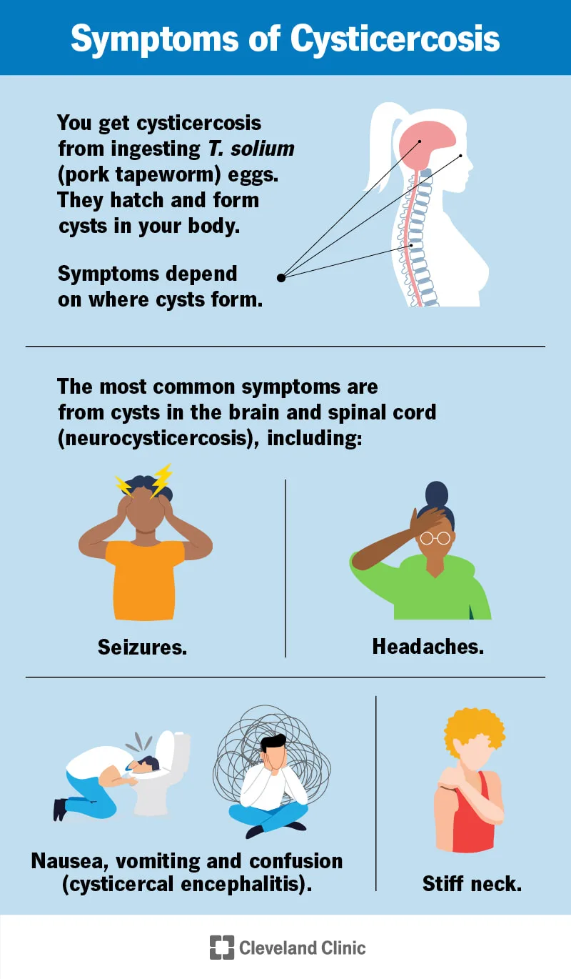
Adult tapeworm infections typically result in minor gastrointestinal symptoms or show no symptoms at all. Cysticercosis is a grave condition that results in the development of cysticerci in many human tissues, such as the eye, brain, muscle, and lungs. The symptoms of cysticerci might differ according on their location, but may encompass meningioencephalitis, seizures, and other neurological signs.
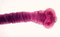
Adult tapeworms remain connected to the intestine through the scolex and can reach a length of up to 5 meters.
Without receiving treatment, they have the potential to live for several decades. In cysticercosis, the eggs are consumed and release oncospheres in the colon, which then go to other tissues in the body.
The presence of proglottids displaying 5-13 uterine branches or eggs in feces or the perianal region is indicative and can be used for diagnosis.
Medical assessment Cysticerci are frequently detectable using X-ray, CT, or MRI imaging due to their tendency to undergo calcification.
Praziquantel is used to treat adult tapeworm infections. The treatment for cysticercosis involves the surgical removal of the cysts and/or the administration of praziquantel or albendazole. Prevention entails refraining from consuming undercooked pork and implementing hygienic practices to prevent the transmission of diseases by the oral ingestion of fecal matter.
Neurological involvement in cysticercosis
Taenia solium is a type of segmented flatworm known as a Cestode. It is commonly referred to as a pork tapeworm.
The strobila consists of one or many hermaphroditic proglottids.
Humans serve as the primary host, while pigs act as the intermediary host. Pigs consume eggs, and the embryo penetrates the intestinal wall, enters the bloodstream, and disseminates to skeletal muscle. Infection in humans occurs when they consume undercooked pork that is contaminated with encysted larvae. Tapeworms mature within the gastrointestinal tract and release proglottids that contain eggs into the feces. The transfer of eggs by the oral route, either from contact with an individual carrying the adult tapeworm or through self-infection, leads to the development of cysticercosis.
Adult tapeworm infections typically result in minor gastrointestinal symptoms or show no symptoms at all. Cysticercosis is a grave condition that results in the development of cysticerci in many human tissues, such as the eye, brain, muscle, and lungs. The symptoms of cysticerci might differ according on their location, but may encompass meningioencephalitis, seizures, and other neurological signs.
Adult tapeworms remain connected to the intestine through the scolex and can reach a length of up to 5 meters.
Without receiving treatment, they have the potential to live for several decades. In cysticercosis, the eggs are consumed and release oncospheres in the colon, which then go to other tissues in the body.
The presence of proglottids displaying 5-13 uterine branches or eggs in feces or the perianal region is indicative and can be used for diagnosis.
Medical assessment Cysticerci are frequently detectable using X-ray, CT, or MRI imaging due to their tendency to undergo calcification.
Praziquantel is used to treat adult tapeworm infections. The treatment for cysticercosis involves the surgical removal of the cysts and/or the administration of praziquantel or albendazole. Prevention entails refraining from consuming undercooked pork and implementing hygienic practices to prevent the transmission of diseases by the oral ingestion of fecal matter.
- Published on
Symptoms and Signs - Differential Diagnosis of Bleeding Time Elevation
• von Willebrand disease
• Uremia
• Thrombocytopenia
• Platelet abnormalities (Bernard-Soulier disease, Glanzmann’s disease)
• Myeloproliferative disorders
• Drugs (aspirin, warfarin, anti-inflammatory medications, streptokinase, urokinase, dextran, b-lactam antibiotics, moxalactam)
• Disseminated intravascular coagulation (DIC)
• Cirrhosis
• Capillary wall abnormalities
• von Willebrand disease
• Uremia
• Thrombocytopenia
• Platelet abnormalities (Bernard-Soulier disease, Glanzmann’s disease)
• Myeloproliferative disorders
• Drugs (aspirin, warfarin, anti-inflammatory medications, streptokinase, urokinase, dextran, b-lactam antibiotics, moxalactam)
• Disseminated intravascular coagulation (DIC)
• Cirrhosis
• Capillary wall abnormalities

- Published on
Symptoms and Signs - Differential Diagnosis of Upper GI Bleeding
• Swallowed hemoptysis
• Oral or pharyngeal lesions: swallowed blood from nose or oropharynx
• Gastric: peptic ulcer (including Cushing’s and Curling’s ulcers), gastritis, angiodysplasia, gastric neoplasms, hiatal hernia, gastric diverticulum, pseudoxanthoma elasticum, Rendu Osler-Weber syndrome
• Esophageal: varices, ulceration, esophagitis, Mallory-Weiss tear, carcinoma, trauma
• Duodenal: peptic ulcer, duodenitis, angiodysplasia, aortoduodenal fistula, duodenal diverticulum, duodenal tumors, carcinoma of ampulla of Vater, parasites (e.g., hookworm), Crohn’s disease
• Biliary: hematobilia (e.g., penetrating injury to liver, hepatobiliary malignant neoplasm, endoscopic papillotomy)
• Swallowed hemoptysis
• Oral or pharyngeal lesions: swallowed blood from nose or oropharynx
• Gastric: peptic ulcer (including Cushing’s and Curling’s ulcers), gastritis, angiodysplasia, gastric neoplasms, hiatal hernia, gastric diverticulum, pseudoxanthoma elasticum, Rendu Osler-Weber syndrome
• Esophageal: varices, ulceration, esophagitis, Mallory-Weiss tear, carcinoma, trauma
• Duodenal: peptic ulcer, duodenitis, angiodysplasia, aortoduodenal fistula, duodenal diverticulum, duodenal tumors, carcinoma of ampulla of Vater, parasites (e.g., hookworm), Crohn’s disease
• Biliary: hematobilia (e.g., penetrating injury to liver, hepatobiliary malignant neoplasm, endoscopic papillotomy)
- Published on
Symptoms and Signs - Differential Diagnosis of Pediatric Upper GI Bleeding
<3 MONTHS
• Swallowed maternal blood
• Gastritis
• Ulcer, stress
• Bleeding diathesis
• Foreign body (nasogastric [NG] tube)
• Vascular malformation
• Duplication
<2 YEARS
• Esophagitis
• Gastritis
• Ulcer
• Pyloric stenosis
• Mallory-Weiss syndrome
• Vascular malformation
• Duplication
<5 YEARS
• Esophagitis
• Gastritis
• Ulcer
• Esophageal varices
• Foreign body
• Mallory-Weiss syndrome
• Hemophilia
• Vascular malformations
5–18 YEARS
• Esophagitis
• Gastritis
• Ulcer
• Esophageal varices
• Mallory-Weiss syndrome
• Inflammatory bowel disease (IBD)
• Hemophilia
• Vascular malformation
<3 MONTHS
• Swallowed maternal blood
• Gastritis
• Ulcer, stress
• Bleeding diathesis
• Foreign body (nasogastric [NG] tube)
• Vascular malformation
• Duplication
<2 YEARS
• Esophagitis
• Gastritis
• Ulcer
• Pyloric stenosis
• Mallory-Weiss syndrome
• Vascular malformation
• Duplication
<5 YEARS
• Esophagitis
• Gastritis
• Ulcer
• Esophageal varices
• Foreign body
• Mallory-Weiss syndrome
• Hemophilia
• Vascular malformations
5–18 YEARS
• Esophagitis
• Gastritis
• Ulcer
• Esophageal varices
• Mallory-Weiss syndrome
• Inflammatory bowel disease (IBD)
• Hemophilia
• Vascular malformation
- Published on
Symptoms and Signs - Differential Diagnosis of Blepharitis
• Phthirus pubis (pubic lice)
• Phthiriasis palpebrarum
• Molluscum contagiosum
• Keratoconjunctivitis sicca
• Herpes simplex blepharitis
• Eyelid malignant neoplasms
• Demodex folliculorum (transparent mites)
• Allergic blepharitis
• Phthirus pubis (pubic lice)
• Phthiriasis palpebrarum
• Molluscum contagiosum
• Keratoconjunctivitis sicca
• Herpes simplex blepharitis
• Eyelid malignant neoplasms
• Demodex folliculorum (transparent mites)
• Allergic blepharitis
- Published on
Symptoms and Signs - Differential Diagnosis of Blindness, Geriatric Age
• Trauma
• Macular degeneration
• Glaucoma
• Diabetic retinopathy
• Corneal scarring
• Cerebrovascular accident (CVA)
• Cataracts
• Trauma
• Macular degeneration
• Glaucoma
• Diabetic retinopathy
• Corneal scarring
• Cerebrovascular accident (CVA)
• Cataracts
- Published on
Symptoms and Signs - Differential Diagnosis of Transient Monocular Blindness
• Psychogenic
• Partial retinal vein occlusion
• Optic disc edema
• Multiple sclerosis
• Migraine (vasospasm)
• Intermittent angle-closure glaucoma
• Hypotension
• Hyphema
• Hypercoagulopathy disorders
• Giant cell arteritis
• Embolic cerebrovascular disease
• Psychogenic
• Partial retinal vein occlusion
• Optic disc edema
• Multiple sclerosis
• Migraine (vasospasm)
• Intermittent angle-closure glaucoma
• Hypotension
• Hyphema
• Hypercoagulopathy disorders
• Giant cell arteritis
• Embolic cerebrovascular disease
