- Published on
Symptoms and Signs - Differential Diagnosis of Blindness, Pediatric Age
CONGENITAL
• Optic nerve hypoplasia or aplasia
• Optic coloboma
• Congenital hydrocephalus
• Hydranencephaly
• Porencephaly
• Microencephaly
• Encephalocele, particularly occipital type
• Morning glory disc
• Aniridia
• Anterior microphthalmia
• Peters’ anomaly
• Persistent pupillary membrane
• Glaucoma
• Cataracts
• Persistent hyperplastic primary vitreous
PHAKOMATOSES
• Tuberous sclerosis
• Neurofibromatosis (special association with optic glioma)
• Sturge-Weber syndrome
• von Hippel–Lindau disease
TUMORS
• Retinoblastoma
• Optic glioma
• Perioptic meningioma
• Craniopharyngioma
• Cerebral glioma
• Posterior and intraventricular tumors when complicated by hydrocephalus
• Pseudotumor cerebri
NEURODEGENERATIVE DISEASES
• Cerebral storage disease
• Gangliosidoses, particularly Tay-Sachs disease (infantile amaurotic familial idiocy), Sandhoff’s variant, generalized gangliosidosis
• Other lipidoses and ceroid lipofuscinoses, particularly the lateonset amaurotic familial idiocies, such as those of Jansky- Bielschowsky disease and of Batten-Mayou disease and
Spielmeyer-Vogt disease
• Mucopolysaccharidoses, particularly Hurler’s syndrome and
Hunter’s syndrome
• Leukodystrophies (dysmyelination disorders), particularly
metachromatic leukodystrophy and Canavan’s disease
• Demyelinating sclerosis (myelinoclastic diseases), especially
Schilder’s disease and Devic’s neuromyelitis optica
• Special types: Dawson’s disease, Leigh disease, Bassen-Kornzweig
syndrome, Refsum’s disease
• Retinal degenerations: retinitis pigmentosa and its variants,
Leber’s congenital type
• Optic atrophies: congenital autosomal recessive type, infantile and
congenital autosomal dominant types, Leber’s disease, and
atrophies associated with hereditary ataxias—the types of Behr, of
Marie, and of Sanger Brown
INFECTIOUS PROCESSES
• Encephalitis, especially in the prenatal infection syndromes caused
by Toxoplasma gondii, cytomegalovirus, rubella virus, Treponema
pallidum, herpes simplex
• Meningitis, arachnoiditis
• Chorioretinitis
• Endophthalmitis
• Keratitis
HEMATOLOGIC DISORDERS
• Leukemia with CNS involvement
VASCULAR AND CIRCULATORY DISORDERS
• Collagen-vascular diseases
• Arteriovenous malformations—intracerebral hemorrhage,
subarachnoid hemorrhage
• Central retinal occlusion
TRAUMA
• Contusion or avulsion of optic nerves, chiasm, globe, cornea
• Cerebral contusion or laceration
• Intracerebral, subarachnoid, or subdural hemorrhage
DRUGS/TOXINS
OTHER
• Retinopathy of prematurity
• Sclerocornea
• Conversion reaction
• Optic neuritis
• Osteopetrosis
CONGENITAL
• Optic nerve hypoplasia or aplasia
• Optic coloboma
• Congenital hydrocephalus
• Hydranencephaly
• Porencephaly
• Microencephaly
• Encephalocele, particularly occipital type
• Morning glory disc
• Aniridia
• Anterior microphthalmia
• Peters’ anomaly
• Persistent pupillary membrane
• Glaucoma
• Cataracts
• Persistent hyperplastic primary vitreous
PHAKOMATOSES
• Tuberous sclerosis
• Neurofibromatosis (special association with optic glioma)
• Sturge-Weber syndrome
• von Hippel–Lindau disease
TUMORS
• Retinoblastoma
• Optic glioma
• Perioptic meningioma
• Craniopharyngioma
• Cerebral glioma
• Posterior and intraventricular tumors when complicated by hydrocephalus
• Pseudotumor cerebri
NEURODEGENERATIVE DISEASES
• Cerebral storage disease
• Gangliosidoses, particularly Tay-Sachs disease (infantile amaurotic familial idiocy), Sandhoff’s variant, generalized gangliosidosis
• Other lipidoses and ceroid lipofuscinoses, particularly the lateonset amaurotic familial idiocies, such as those of Jansky- Bielschowsky disease and of Batten-Mayou disease and
Spielmeyer-Vogt disease
• Mucopolysaccharidoses, particularly Hurler’s syndrome and
Hunter’s syndrome
• Leukodystrophies (dysmyelination disorders), particularly
metachromatic leukodystrophy and Canavan’s disease
• Demyelinating sclerosis (myelinoclastic diseases), especially
Schilder’s disease and Devic’s neuromyelitis optica
• Special types: Dawson’s disease, Leigh disease, Bassen-Kornzweig
syndrome, Refsum’s disease
• Retinal degenerations: retinitis pigmentosa and its variants,
Leber’s congenital type
• Optic atrophies: congenital autosomal recessive type, infantile and
congenital autosomal dominant types, Leber’s disease, and
atrophies associated with hereditary ataxias—the types of Behr, of
Marie, and of Sanger Brown
INFECTIOUS PROCESSES
• Encephalitis, especially in the prenatal infection syndromes caused
by Toxoplasma gondii, cytomegalovirus, rubella virus, Treponema
pallidum, herpes simplex
• Meningitis, arachnoiditis
• Chorioretinitis
• Endophthalmitis
• Keratitis
HEMATOLOGIC DISORDERS
• Leukemia with CNS involvement
VASCULAR AND CIRCULATORY DISORDERS
• Collagen-vascular diseases
• Arteriovenous malformations—intracerebral hemorrhage,
subarachnoid hemorrhage
• Central retinal occlusion
TRAUMA
• Contusion or avulsion of optic nerves, chiasm, globe, cornea
• Cerebral contusion or laceration
• Intracerebral, subarachnoid, or subdural hemorrhage
DRUGS/TOXINS
OTHER
• Retinopathy of prematurity
• Sclerocornea
• Conversion reaction
• Optic neuritis
• Osteopetrosis

- Published on
Symptoms and Signs - Differential Diagnosis of Blisters, Subepidermal
• Variegate porphyria
• Urticaria pigmentosa
• Toxic epidermal necrosis
• Pseudoporphyria
• Pressure necrosis
• Porphyria cutanea tarda
• Polymorphous light eruption
• Lupus erythematosus
• Linear IgA disease
• Leukocytoclastic vasculitis
• Epidermolysis bullosa
• Dermatitis herpetiformis
• Burns
• Bullous pemphigoid
• Bullous drug reaction
• Arthropod bite reaction
• Amyloidosis
• Acute graft-versus-host reaction
• Variegate porphyria
• Urticaria pigmentosa
• Toxic epidermal necrosis
• Pseudoporphyria
• Pressure necrosis
• Porphyria cutanea tarda
• Polymorphous light eruption
• Lupus erythematosus
• Linear IgA disease
• Leukocytoclastic vasculitis
• Epidermolysis bullosa
• Dermatitis herpetiformis
• Burns
• Bullous pemphigoid
• Bullous drug reaction
• Arthropod bite reaction
• Amyloidosis
• Acute graft-versus-host reaction
- Published on
Symptoms and Signs - Differential Diagnosis of Body Dysmorphic Disorder
• Social phobia
• Personality disorder
• Obsessive-compulsive disorder
• Hypochondriasis
• Anxiety disorder
• Anorexia nervosa
• Social phobia
• Personality disorder
• Obsessive-compulsive disorder
• Hypochondriasis
• Anxiety disorder
• Anorexia nervosa
- Published on
Symptoms and Signs - Differential Diagnosis of Bone Lesions, Preferential Site of Origin
EPIPHYSIS
• Chondroblastoma
• Giant cell tumor—after fusion of growth plate
• Langerhans cell histiocytosis
• Clear cell chondrosarcoma
• Osteosarcoma
METAPHYSIS
• Parosteal sarcoma
• Chondrosarcoma
• Fibrosarcoma
• Nonossifying fibroma
• Giant cell tumor—before fusion of growth plate
• Unicameral bone cyst
• Aneurysmal bone cyst
DIAPHYSIS
• Myeloma
• Ewing’s tumor
• Reticulum cell sarcoma
METADIAPHYSEAL
• Fibrosarcoma
• Fibrous dysplasia
• Enchondroma
• Osteoid osteoma
• Chondromyofibroma
EPIPHYSIS
• Chondroblastoma
• Giant cell tumor—after fusion of growth plate
• Langerhans cell histiocytosis
• Clear cell chondrosarcoma
• Osteosarcoma
METAPHYSIS
• Parosteal sarcoma
• Chondrosarcoma
• Fibrosarcoma
• Nonossifying fibroma
• Giant cell tumor—before fusion of growth plate
• Unicameral bone cyst
• Aneurysmal bone cyst
DIAPHYSIS
• Myeloma
• Ewing’s tumor
• Reticulum cell sarcoma
METADIAPHYSEAL
• Fibrosarcoma
• Fibrous dysplasia
• Enchondroma
• Osteoid osteoma
• Chondromyofibroma
- Published on
Infectious Diseases and Microbiology - Trichuriasis / Whipworm Infection ( Trichuris trichiura)
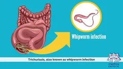
Whipworm infection, also known as trichuriasis
The Trichuris trichiura is a nematode, also known as a roundworm. The term "whipworm" is also used.
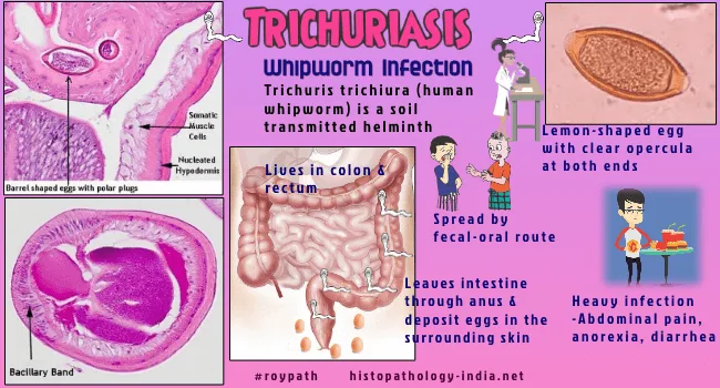
Adult worms have an anterior end that resembles a thread and is embedded in the mucosa of the large intestine when they are fully mature.
Ingestion of eggs that have been infected with human excrement is the means by which the disease is transmitted. Every day, adult females generate between three thousand and ten thousand eggs, which are then expelled through the feces. The incubation period of at least ten days in the soil is required for them to become infectious. There are regions in the rural southeast of the United States that are affected by the parasite, which has a global spread. Utilizing human feces as a fertilizer is a factor that promotes to the spread of infection.
Depending on the magnitude of the worm infestation, clinical symptoms can differ. It is common for minor loads to be asymptomatic. The presence of greater worm loads is associated with the development of symptoms such as abdominal pain, bloody diarrhea, tenesmus, rectal prolapse, anemia, and eosinophilia, in addition to a loss of nutritional supplements and weight. The loss of a substantial amount of blood is possible in severe situations.
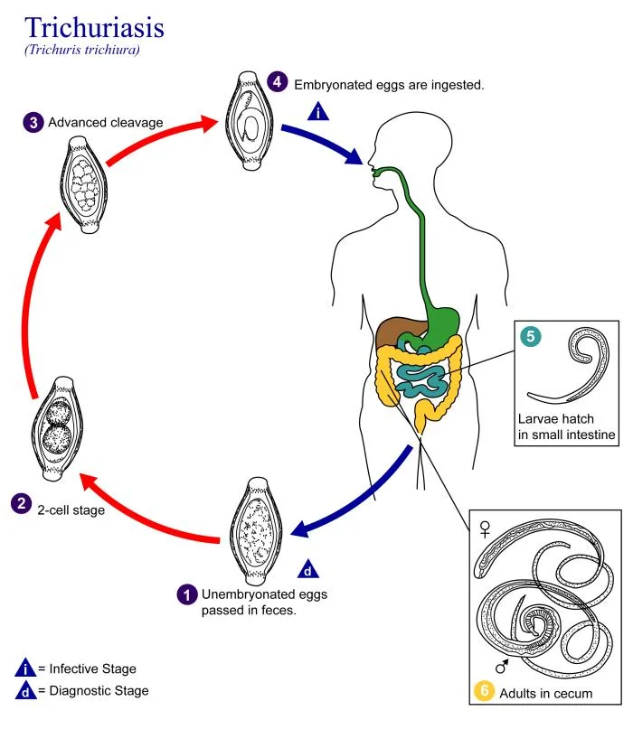
After the eggs have been consumed, the larvae will hatch in the small intestine and then be transported to the colon, where they will eventually develop into adults. On the colon, the adult forms adhere themselves, which results in local ulceration. They have a lifespan of four to eight years.
On direct examination of the feces used for diagnosis or through the application of concentration techniques, one can discover eggs that have the distinctive shape of a barrel and have a plug on each end.
Mebendazole or albendazole are both potential treatments for the condition. Proper disposal of human feces and maintaining a high level of personal hygiene are also essential components of prevention techniques.
Whipworm infection, also known as trichuriasis
The Trichuris trichiura is a nematode, also known as a roundworm. The term "whipworm" is also used.
Adult worms have an anterior end that resembles a thread and is embedded in the mucosa of the large intestine when they are fully mature.
Ingestion of eggs that have been infected with human excrement is the means by which the disease is transmitted. Every day, adult females generate between three thousand and ten thousand eggs, which are then expelled through the feces. The incubation period of at least ten days in the soil is required for them to become infectious. There are regions in the rural southeast of the United States that are affected by the parasite, which has a global spread. Utilizing human feces as a fertilizer is a factor that promotes to the spread of infection.
Depending on the magnitude of the worm infestation, clinical symptoms can differ. It is common for minor loads to be asymptomatic. The presence of greater worm loads is associated with the development of symptoms such as abdominal pain, bloody diarrhea, tenesmus, rectal prolapse, anemia, and eosinophilia, in addition to a loss of nutritional supplements and weight. The loss of a substantial amount of blood is possible in severe situations.
After the eggs have been consumed, the larvae will hatch in the small intestine and then be transported to the colon, where they will eventually develop into adults. On the colon, the adult forms adhere themselves, which results in local ulceration. They have a lifespan of four to eight years.
On direct examination of the feces used for diagnosis or through the application of concentration techniques, one can discover eggs that have the distinctive shape of a barrel and have a plug on each end.
Mebendazole or albendazole are both potential treatments for the condition. Proper disposal of human feces and maintaining a high level of personal hygiene are also essential components of prevention techniques.
- Published on
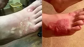
Infectious Diseases and Microbiology - HookwormInfection (Ancylostoma duodenale and Necator americanus)
Hookworm Infection
Both Necator americanus and Ancylostoma duodenale. These are both roundworms, or nematodes, and they lead similar lives.
Likewise known as hookworm. The primary reservoir is people.
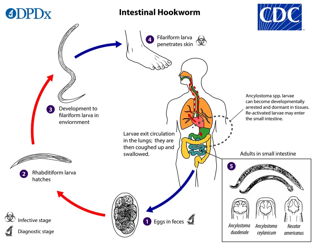
In the soil, eggs hatch into rhabditoid larvae, which then transform into infectious filariform larvae that can live there for several weeks or months. Filariform larvae directly penetrate the skin to cause infection. Larvae travel through the bloodstream to the heart and lungs. Eventually, they ascend the pulmonary tree, are swallowed, and arrive in the small intestine, where they mature into adult worms with a daily egg production of at least 5,000 and a lifespan of five to fifteen years. The cycle is then completed when the eggs hatch, pollute the soil, and are passed on in feces.
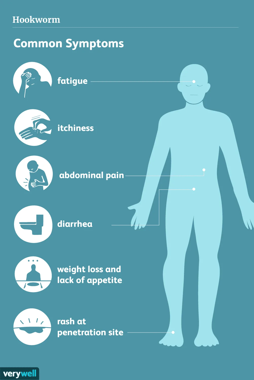
Most people don't have any symptoms, but others may experience pneumonitis during the larval lung migration or an allergic rash at the point of entry. Excessive worm loads can cause exhaustion, weight loss, and mental or physical impairment. Microcytic hypochromic anemia is brought on by a persistent infection.
Worm burden is directly correlated with pathogenesis. Because adult worms are blood-feeders, blood loss of up to 0.2 mL per worm each day and anemia may arise. Lung migration in larvae is linked to eosinophilia and pneumonia.
The diagnosis is made by showing hookworm eggs in feces under a microscope.
Worms can be killed with albendazole, mebendazole, and pyrantel pamoate. Wearing shoes, learning about transmission, and disposing of human waste properly are all part of prevention.
Hookworm Infection
Both Necator americanus and Ancylostoma duodenale. These are both roundworms, or nematodes, and they lead similar lives.
Likewise known as hookworm. The primary reservoir is people.
In the soil, eggs hatch into rhabditoid larvae, which then transform into infectious filariform larvae that can live there for several weeks or months. Filariform larvae directly penetrate the skin to cause infection. Larvae travel through the bloodstream to the heart and lungs. Eventually, they ascend the pulmonary tree, are swallowed, and arrive in the small intestine, where they mature into adult worms with a daily egg production of at least 5,000 and a lifespan of five to fifteen years. The cycle is then completed when the eggs hatch, pollute the soil, and are passed on in feces.
Most people don't have any symptoms, but others may experience pneumonitis during the larval lung migration or an allergic rash at the point of entry. Excessive worm loads can cause exhaustion, weight loss, and mental or physical impairment. Microcytic hypochromic anemia is brought on by a persistent infection.
Worm burden is directly correlated with pathogenesis. Because adult worms are blood-feeders, blood loss of up to 0.2 mL per worm each day and anemia may arise. Lung migration in larvae is linked to eosinophilia and pneumonia.
The diagnosis is made by showing hookworm eggs in feces under a microscope.
Worms can be killed with albendazole, mebendazole, and pyrantel pamoate. Wearing shoes, learning about transmission, and disposing of human waste properly are all part of prevention.
- Published on
Infectious Diseases and Microbiology - Strongyloidiasis ( Strongyloides stercoralis)
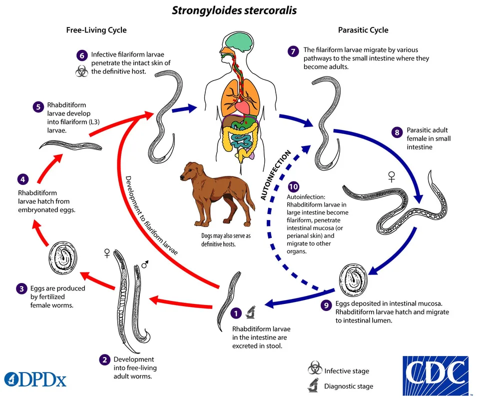
Strongyloidiasis is a parasitic infection which is caused by Strongyloides stercoralis which is a parasitic nematode.
An intestinal nematode, also known as a roundworm. Distinguished by its ability to alternate between a parasitic life cycle and a free-living cycle.
Threadworm is an alternative name for this organism.
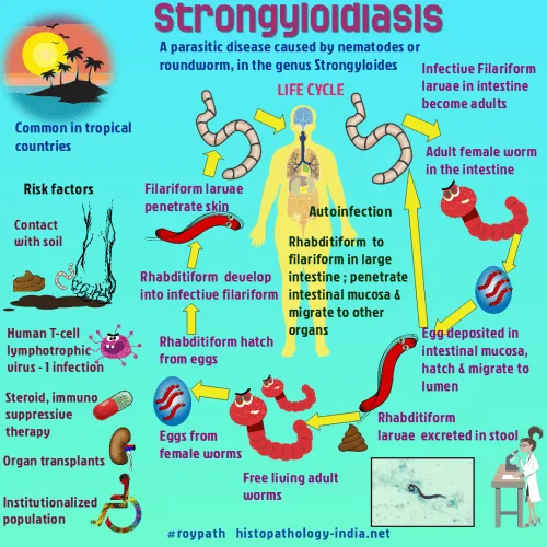
Located throughout the southern and southwestern regions of the United States, as well as in tropical regions across the globe. Infection occurs when fillariform larvae in soil infected with human feces penetrate the skin directly, similar to hookworm infection. Eggs excreted in feces have the potential to mature into larvae and subsequently adults in the soil, giving rise to eggs and larvae outside the host.
Eosinophilia can occur as the sole indication of an infection. Symptomatic infections may manifest as pnemonitis, watery diarrhea, abdominal pain, and the presence of blood in the stool, depending on the worm burden and immunologic competency of the individual. Hyperinfection syndrome, which occurs in persons with weakened immune systems (such as those on corticosteroids), is characterized by the spread of larvae to several organs, including the lungs, central nervous system, and heart.
Perforation of the colonic wall might result in subsequent bacterial infections. Autoinfection is frequently observed in individuals who have a large number of worms in their body.
Following entrance, the larvae undergo a process of lung migration. The maturation of worms occurs within the small intestine, where they subsequently generate eggs. Ova have the ability to hatch within the intestinal tract and the developing larvae can directly infiltrate the intestine, leading to internal autoinfection.
Larvae that infect the region around the anus might lead to external autoinfection. Additional larvae are excreted in feces.
The development of disease depends on the number of worms present.
Feces testing may reveal distinctive larvae, but it may be necessary to check multiple fresh feces samples before a definitive diagnosis is made.
Ivermectin, thiabendazole, or albendazole are effective treatments for the majority of patients. Preventive measures encompass the utilization of footwear, dissemination of knowledge regarding transmission and hygiene, and the management of human excrement.
Strongyloidiasis is a parasitic infection which is caused by Strongyloides stercoralis which is a parasitic nematode.
An intestinal nematode, also known as a roundworm. Distinguished by its ability to alternate between a parasitic life cycle and a free-living cycle.
Threadworm is an alternative name for this organism.
Located throughout the southern and southwestern regions of the United States, as well as in tropical regions across the globe. Infection occurs when fillariform larvae in soil infected with human feces penetrate the skin directly, similar to hookworm infection. Eggs excreted in feces have the potential to mature into larvae and subsequently adults in the soil, giving rise to eggs and larvae outside the host.
Eosinophilia can occur as the sole indication of an infection. Symptomatic infections may manifest as pnemonitis, watery diarrhea, abdominal pain, and the presence of blood in the stool, depending on the worm burden and immunologic competency of the individual. Hyperinfection syndrome, which occurs in persons with weakened immune systems (such as those on corticosteroids), is characterized by the spread of larvae to several organs, including the lungs, central nervous system, and heart.
Perforation of the colonic wall might result in subsequent bacterial infections. Autoinfection is frequently observed in individuals who have a large number of worms in their body.
Following entrance, the larvae undergo a process of lung migration. The maturation of worms occurs within the small intestine, where they subsequently generate eggs. Ova have the ability to hatch within the intestinal tract and the developing larvae can directly infiltrate the intestine, leading to internal autoinfection.
Larvae that infect the region around the anus might lead to external autoinfection. Additional larvae are excreted in feces.
The development of disease depends on the number of worms present.
Feces testing may reveal distinctive larvae, but it may be necessary to check multiple fresh feces samples before a definitive diagnosis is made.
Ivermectin, thiabendazole, or albendazole are effective treatments for the majority of patients. Preventive measures encompass the utilization of footwear, dissemination of knowledge regarding transmission and hygiene, and the management of human excrement.
- Published on
Infectious Diseases and Microbiology - Trichinosis (Trichinella spiralis)
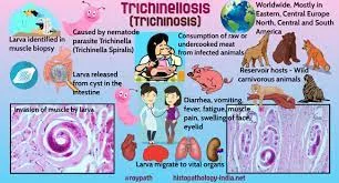
Trichinosis is a disease caused by the roundworm Trichinella spiralis, which belongs to the Nematode family. Gastrointestinal and cellular infections.
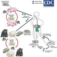
The transmission occurs when individuals consume undercooked pork, bear, seal, or deer meat that contains encysted T. spiralis in the muscle tissue. Humans are an evolutionary dead-end as a host.
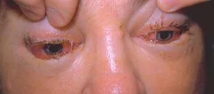
The clinical manifestation of Trichinella spiralis results in the development of trichinosis. The intestinal stage is marked by gastrointestinal symptoms such as abdominal discomfort, diarrhea, nausea, and vomiting. The tissue stage typically manifests several weeks after the onset of gastrointestinal symptoms. It may present with many symptoms such as rash, fever, myalgia, conjunctival and subungual bleeding, splinter hemorrhages, periorbital edema, central nervous system problems, congestive heart failure, and respiratory arrest. The symptoms observed during the tissue stage of the infection are contingent upon the specific location and quantity of the larvae in migration.
The act of consuming insufficiently cooked muscle tissue that contains larvae enclosed in cysts leads to the digestion of the larvae. The larvae then travel to the small intestine and penetrate the epithelium, where they mature into adults. They subsequently return to the intestinal lumen and engage in reproduction. Adult females produce embryonated eggs within their uterus. The larvae generated infiltrate the intestinal wall and travel through the bloodstream to several organs. Subsequently, they form cysts within the skeletal muscle. The development of the disease is caused by an inflammatory response involving eosinophils, which is influenced by the quantity of migratory larvae and cysts.
The diagnosis of eosinophilia involves the finding of larvae in striated muscle from biopsy specimens, as well as the use of serology.
Albendazole or mebendazole have been employed to eradicate mature worms. There are currently no recognized techniques to eliminate encysted larvae. Corticosteroids have the ability to mitigate inflammation. Preventive measures involve minimizing the transfer of diseases to pigs by refraining from feeding them with waste materials and abstaining from consuming foods that are not thoroughly prepared.
Trichinosis is a disease caused by the roundworm Trichinella spiralis, which belongs to the Nematode family. Gastrointestinal and cellular infections.
The transmission occurs when individuals consume undercooked pork, bear, seal, or deer meat that contains encysted T. spiralis in the muscle tissue. Humans are an evolutionary dead-end as a host.
The clinical manifestation of Trichinella spiralis results in the development of trichinosis. The intestinal stage is marked by gastrointestinal symptoms such as abdominal discomfort, diarrhea, nausea, and vomiting. The tissue stage typically manifests several weeks after the onset of gastrointestinal symptoms. It may present with many symptoms such as rash, fever, myalgia, conjunctival and subungual bleeding, splinter hemorrhages, periorbital edema, central nervous system problems, congestive heart failure, and respiratory arrest. The symptoms observed during the tissue stage of the infection are contingent upon the specific location and quantity of the larvae in migration.
The act of consuming insufficiently cooked muscle tissue that contains larvae enclosed in cysts leads to the digestion of the larvae. The larvae then travel to the small intestine and penetrate the epithelium, where they mature into adults. They subsequently return to the intestinal lumen and engage in reproduction. Adult females produce embryonated eggs within their uterus. The larvae generated infiltrate the intestinal wall and travel through the bloodstream to several organs. Subsequently, they form cysts within the skeletal muscle. The development of the disease is caused by an inflammatory response involving eosinophils, which is influenced by the quantity of migratory larvae and cysts.
The diagnosis of eosinophilia involves the finding of larvae in striated muscle from biopsy specimens, as well as the use of serology.
Albendazole or mebendazole have been employed to eradicate mature worms. There are currently no recognized techniques to eliminate encysted larvae. Corticosteroids have the ability to mitigate inflammation. Preventive measures involve minimizing the transfer of diseases to pigs by refraining from feeding them with waste materials and abstaining from consuming foods that are not thoroughly prepared.
- Published on
Infectious Diseases and Microbiology - Filariasis (Wuchereria bancrofti)
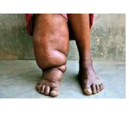
Lymphatic obstruction caused by filariasis.
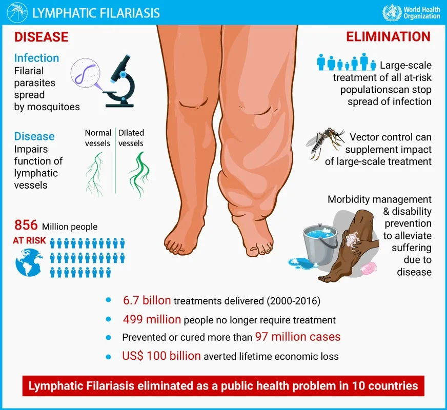
Wuchereria bancrofti, together with Brugia malayi and B timori, are nematodes that reside in the tissues and belong to the filarial group of roundworms.
Transmitted by an arthropod carrier.The transmission of W bancrofti can be facilitated by several mosquito genera, including Anopheles, Culex, Aedes, and Mansonia.
W bancrofti is prevalent in numerous regions in Africa, Asia, South America, and the southern Mediterranean.The majority of filarial infections do not exhibit any symptoms. The initial signs of the condition include fever, chills, muscle pain, and lymphadenitis. Chronic inflammation causes blockage of the lymphatic system, which can lead to elephantiasis in extreme instances. Additionally, secondary bacterial infections may arise.
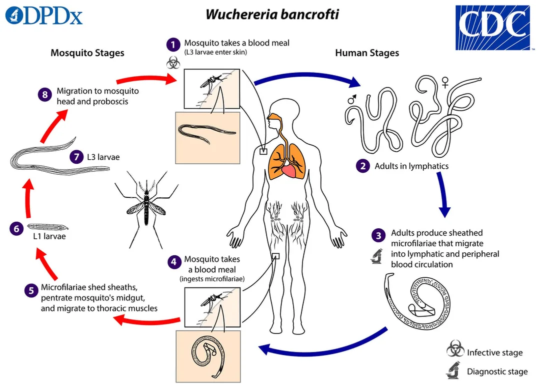
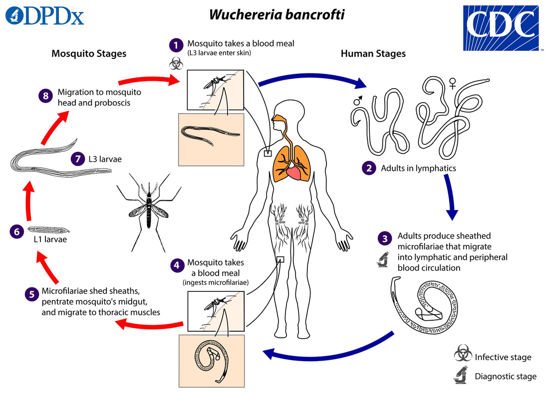
After being bitten by an infected mosquito, the larvae enter the skin and travel through the lymphatic system to the nearby lymph nodes. Following an extended period of incubation lasting between 6 months and a year, the larvae undergo maturation and transform into adult organisms. These adults then generate microfilarial larvae which travel into the bloodstream during nighttime. The microfilariae can be consumed by a feeding mosquito to continue the transmission process. The adult versions have the ability to endure for a period of up to ten years. Chronic disease arises due to inflammation in the lymphatic system, leading to blockage and swelling. Recurring infections escalate the number of worms present, resulting in progressively severe symptoms of the condition.
The microfilariae undergo nocturnal migration into the bloodstream. The thick-smear blood examination should be conducted using samples collected specifically between the hours of 10 pm and 4 am.
Diethylcarbamazine is employed for the eradication of adult worms and microfilariae. Obstructions may require surgical intervention for relief.
Prevention entails implementing measures to control mosquitoes, as well as employing personal barriers and repellents to minimize exposure. In recent times, a strategy known as community-wide treatment has been implemented to mitigate the spread of diseases in places with a high risk of transmission.
Lymphatic obstruction caused by filariasis.
Wuchereria bancrofti, together with Brugia malayi and B timori, are nematodes that reside in the tissues and belong to the filarial group of roundworms.
Transmitted by an arthropod carrier.The transmission of W bancrofti can be facilitated by several mosquito genera, including Anopheles, Culex, Aedes, and Mansonia.
W bancrofti is prevalent in numerous regions in Africa, Asia, South America, and the southern Mediterranean.The majority of filarial infections do not exhibit any symptoms. The initial signs of the condition include fever, chills, muscle pain, and lymphadenitis. Chronic inflammation causes blockage of the lymphatic system, which can lead to elephantiasis in extreme instances. Additionally, secondary bacterial infections may arise.
After being bitten by an infected mosquito, the larvae enter the skin and travel through the lymphatic system to the nearby lymph nodes. Following an extended period of incubation lasting between 6 months and a year, the larvae undergo maturation and transform into adult organisms. These adults then generate microfilarial larvae which travel into the bloodstream during nighttime. The microfilariae can be consumed by a feeding mosquito to continue the transmission process. The adult versions have the ability to endure for a period of up to ten years. Chronic disease arises due to inflammation in the lymphatic system, leading to blockage and swelling. Recurring infections escalate the number of worms present, resulting in progressively severe symptoms of the condition.
The microfilariae undergo nocturnal migration into the bloodstream. The thick-smear blood examination should be conducted using samples collected specifically between the hours of 10 pm and 4 am.
Diethylcarbamazine is employed for the eradication of adult worms and microfilariae. Obstructions may require surgical intervention for relief.
Prevention entails implementing measures to control mosquitoes, as well as employing personal barriers and repellents to minimize exposure. In recent times, a strategy known as community-wide treatment has been implemented to mitigate the spread of diseases in places with a high risk of transmission.
- Published on
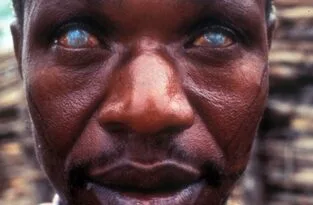
Infectious Diseases and Microbiology - Onchocerciasis/ River Blindness (Onchocerca volvulus )
Onchocerciasis, often known as River Blindness, is a parasitic disease.
Onchocerca volvulus is a parasitic roundworm belonging to the nematode family.
Transmitted by the intermediary of an arthropod vector.
The Simulium black fly, which reproduces in swiftly moving streams and rivers, serves as the carrier of the disease.
Geographical regions where a certain disease is regularly seen include equatorial Africa and Central and South America.
The clinical causative agent of river blindness. The symptoms encompass a skin rash, itching, reduced elasticity in the subcutaneous tissue (resulting in the formation of enormous skin folds), and varying degrees of visual impairment, ranging from partial to complete blindness.
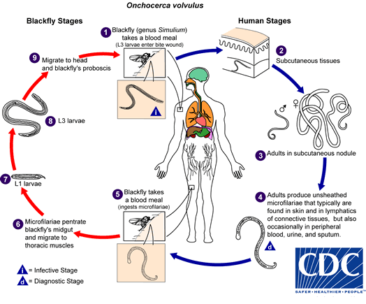
Upon entering the bite wound, the larvae proceed to travel towards the subcutaneous tissue, where they undergo development and transform into adults within fibrous nodules located beneath the skin. Once the worms reach maturity, they create microfilariae that travel through the subcutaneous tissue and skin. These microfilariae can be consumed by biting black flies, which helps to spread the infection.
The pathogenesis occurs due to an exaggerated immune response to the antigens of the parasite and the inflammation caused by the movement of the larvae. Microfilariae in ocular structures cause photophobia and inflammation in several parts of the eye, including the cornea, iris, ciliary body, retina, choroids, and optic nerve. Untreated illness can lead to blindness.
Microfilariae are observable in skin biopsies and can also be seen through slit-lamp examination of the anterior chamber of an affected eye.
The treatment consists of surgically extracting mature cutaneous nodules along with the administration of ivermectin (to eliminate microfilariae). Prevention encompasses the implementation of infection control strategies. Administering prophylactic ivermectin treatment in places where a disease is prevalent can decrease the number of microfilariae present.
Onchocerciasis, often known as River Blindness, is a parasitic disease.
Onchocerca volvulus is a parasitic roundworm belonging to the nematode family.
Transmitted by the intermediary of an arthropod vector.
The Simulium black fly, which reproduces in swiftly moving streams and rivers, serves as the carrier of the disease.
Geographical regions where a certain disease is regularly seen include equatorial Africa and Central and South America.
The clinical causative agent of river blindness. The symptoms encompass a skin rash, itching, reduced elasticity in the subcutaneous tissue (resulting in the formation of enormous skin folds), and varying degrees of visual impairment, ranging from partial to complete blindness.
Upon entering the bite wound, the larvae proceed to travel towards the subcutaneous tissue, where they undergo development and transform into adults within fibrous nodules located beneath the skin. Once the worms reach maturity, they create microfilariae that travel through the subcutaneous tissue and skin. These microfilariae can be consumed by biting black flies, which helps to spread the infection.
The pathogenesis occurs due to an exaggerated immune response to the antigens of the parasite and the inflammation caused by the movement of the larvae. Microfilariae in ocular structures cause photophobia and inflammation in several parts of the eye, including the cornea, iris, ciliary body, retina, choroids, and optic nerve. Untreated illness can lead to blindness.
Microfilariae are observable in skin biopsies and can also be seen through slit-lamp examination of the anterior chamber of an affected eye.
The treatment consists of surgically extracting mature cutaneous nodules along with the administration of ivermectin (to eliminate microfilariae). Prevention encompasses the implementation of infection control strategies. Administering prophylactic ivermectin treatment in places where a disease is prevalent can decrease the number of microfilariae present.