- Published on
Infectious Diseases and Microbiology - American Trypanosomiasis ( Trypanosoma cruzi)
Trypanosoma cruzi is a parasitic organism that resides in the blood and tissues and possesses a whip-like structure called a flagellum.
Tissue morphology: amastigote (lacking flagella). Blood morphology: trypomastigote (possessing a flagellum).
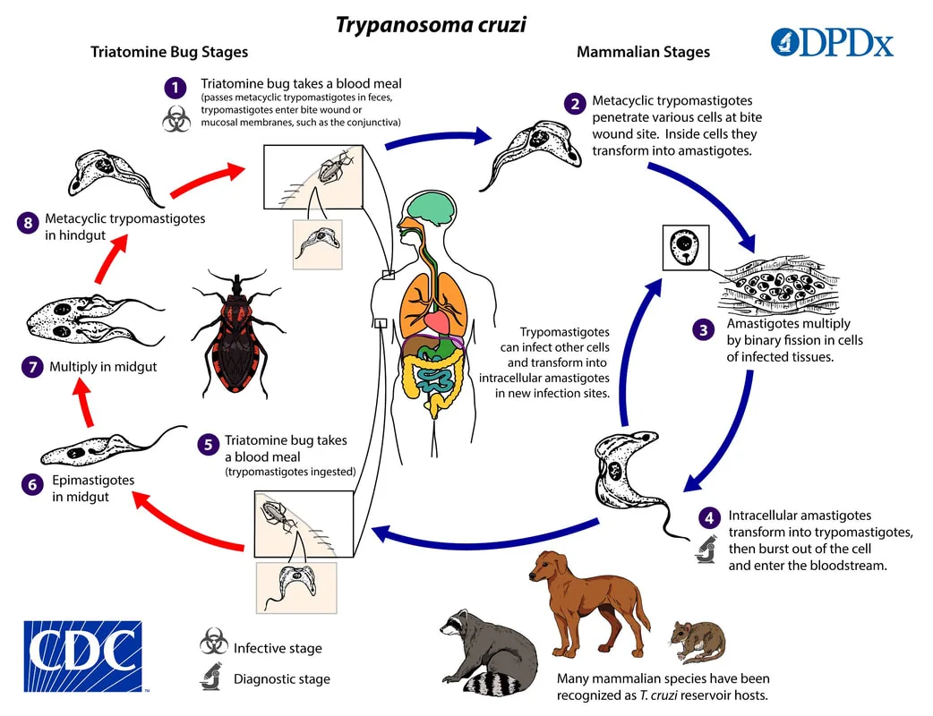
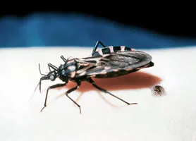
Mammals and humans act as reservoirs. The vector is the Triatoma insect, sometimes known as the "kissing bug". The infective form is introduced when the feces of an infected bug, which are deposited during feeding, are rubbed into the conjunctiva, the bite site, or a skin break. The prevalence of the disease is primarily observed in Mexico, as well as in Central and South America.
The clinical manifestation of T cruzi infection results in American trypanosomiasis, also known as Chagas disease. This condition is defined by the presence of an erythematous chagoma, which appears on the face or arms at the site of the insect bite. In cases when the infection occurs in the eye, symptoms such as edema, conjunctivitis, and the development of local lymphadenopathy, known as the Romaña sign, may occur. The disease is marked by symptoms such as fever, chills, muscle pain, swollen lymph nodes, and enlargement of the liver and spleen. In severe cases, it can lead to complications such as inflammation of the brain and spinal cord, inflammation of the heart muscle, enlargement of the esophagus, and enlargement of the colon.
After entering the host, trypomastigotes travel through the bloodstream and have the ability to infect various tissues, such as muscle, heart, and glial cells. Upon entering the cell, the amastigote form undergoes development, replication, and ultimately causes the demise of the cell, leading to the liberation of parasites capable of infiltrating further cells.
Trypomastigote forms can be identified in both thick and thin blood films during the acute phase of the disease. For persistent infections, which have a low level of parasites in the blood, it is recommended to recover the parasites by growing them on a specific medium or by identifying them by xenodiagnosis. Xenodiagnosis involves allowing a non-infected reduviid bug to feed on the patient and get infected.
The acute phase of the disease can be managed using either nifurtimox or benznidazole. Prevention entails refraining from coming into contact with reduviid insects.
Trypanosoma cruzi is a parasitic organism that resides in the blood and tissues and possesses a whip-like structure called a flagellum.
Tissue morphology: amastigote (lacking flagella). Blood morphology: trypomastigote (possessing a flagellum).
Mammals and humans act as reservoirs. The vector is the Triatoma insect, sometimes known as the "kissing bug". The infective form is introduced when the feces of an infected bug, which are deposited during feeding, are rubbed into the conjunctiva, the bite site, or a skin break. The prevalence of the disease is primarily observed in Mexico, as well as in Central and South America.
The clinical manifestation of T cruzi infection results in American trypanosomiasis, also known as Chagas disease. This condition is defined by the presence of an erythematous chagoma, which appears on the face or arms at the site of the insect bite. In cases when the infection occurs in the eye, symptoms such as edema, conjunctivitis, and the development of local lymphadenopathy, known as the Romaña sign, may occur. The disease is marked by symptoms such as fever, chills, muscle pain, swollen lymph nodes, and enlargement of the liver and spleen. In severe cases, it can lead to complications such as inflammation of the brain and spinal cord, inflammation of the heart muscle, enlargement of the esophagus, and enlargement of the colon.
After entering the host, trypomastigotes travel through the bloodstream and have the ability to infect various tissues, such as muscle, heart, and glial cells. Upon entering the cell, the amastigote form undergoes development, replication, and ultimately causes the demise of the cell, leading to the liberation of parasites capable of infiltrating further cells.
Trypomastigote forms can be identified in both thick and thin blood films during the acute phase of the disease. For persistent infections, which have a low level of parasites in the blood, it is recommended to recover the parasites by growing them on a specific medium or by identifying them by xenodiagnosis. Xenodiagnosis involves allowing a non-infected reduviid bug to feed on the patient and get infected.
The acute phase of the disease can be managed using either nifurtimox or benznidazole. Prevention entails refraining from coming into contact with reduviid insects.
- Published on
Infectious Diseases and Microbiology - Malaria ( Plasmodium species)
Plasmodium species causing malaria.It is a sporozoan parasite that undergoes alternating cycles of sexual and asexual reproduction. The species that cause infections in humans are P vivax, P ovale, P malariae, and P falciparum.
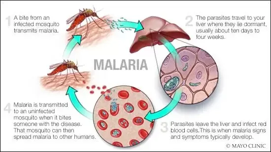
The female Anopheles mosquito serves as both the definitive host and the vector for transmission. The sexual cycle, known as sporogony, takes place within the mosquito host, while the asexual stage, referred to as schizogony, occurs within the human intermediate host. P. vivax and P. ovale have the ability to develop hypnozoites in the liver and remain in a dormant state for an extended period of time.
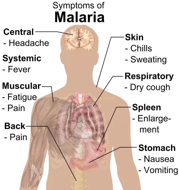
Malaria is a recurring illness that progresses through three distinct stages. The "cold stage" is characterized by shaking chills, the "hot stage" is characterized by fevers as high as 41°C, and the "sweating stage" is characterized by drenching sweats and weariness.
Additional symptoms include enlargement of the spleen, enlargement of the liver, and a decrease in red blood cells. P. falciparum is responsible for causing severe illness, which includes the blockage of small blood vessels, damage to the brain and kidneys, acute kidney failure, and ultimately, death.
The primary infection attacks hepatocytes. Hepatocyte schizonts liberate many merozoites into the circulatory system.
P. vivax and P. ovale specifically invade reticulocytes, while P. malariae infects mature erythrocytes. P. falciparum, on the other hand, has the ability to infect all types of red blood cells (RBCs). The red blood cells that are infected undergo the formation of a ring-shaped trophozoite, which then develops into a schizont containing 8-24 merozoites. Rupture leads to episodes of fever.
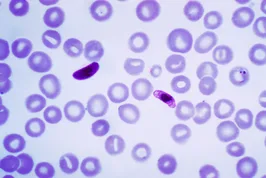
Every species exhibits distinctive microscopic features that are observable in both thick and thin blood smears.
Chloroquine demonstrates efficacy in the erythrocytic phases. Primaquine eradicates dormant liver stages and gametocytes of the malaria parasite P. falciparum for preventive purposes. Nevertheless, the prevalence of chloroquine resistance is high. Other options are Malarone, quinine, quinidine, mefloquine, and pyrimethamine-sulfadoxine. Prevention entails the use of prophylaxis, such as chloroquine or other antimalarial medications, together with bed nets that are treated with pesticide and mosquito repellents.
Plasmodium species causing malaria.It is a sporozoan parasite that undergoes alternating cycles of sexual and asexual reproduction. The species that cause infections in humans are P vivax, P ovale, P malariae, and P falciparum.
The female Anopheles mosquito serves as both the definitive host and the vector for transmission. The sexual cycle, known as sporogony, takes place within the mosquito host, while the asexual stage, referred to as schizogony, occurs within the human intermediate host. P. vivax and P. ovale have the ability to develop hypnozoites in the liver and remain in a dormant state for an extended period of time.
Malaria is a recurring illness that progresses through three distinct stages. The "cold stage" is characterized by shaking chills, the "hot stage" is characterized by fevers as high as 41°C, and the "sweating stage" is characterized by drenching sweats and weariness.
Additional symptoms include enlargement of the spleen, enlargement of the liver, and a decrease in red blood cells. P. falciparum is responsible for causing severe illness, which includes the blockage of small blood vessels, damage to the brain and kidneys, acute kidney failure, and ultimately, death.
The primary infection attacks hepatocytes. Hepatocyte schizonts liberate many merozoites into the circulatory system.
P. vivax and P. ovale specifically invade reticulocytes, while P. malariae infects mature erythrocytes. P. falciparum, on the other hand, has the ability to infect all types of red blood cells (RBCs). The red blood cells that are infected undergo the formation of a ring-shaped trophozoite, which then develops into a schizont containing 8-24 merozoites. Rupture leads to episodes of fever.
Every species exhibits distinctive microscopic features that are observable in both thick and thin blood smears.
Chloroquine demonstrates efficacy in the erythrocytic phases. Primaquine eradicates dormant liver stages and gametocytes of the malaria parasite P. falciparum for preventive purposes. Nevertheless, the prevalence of chloroquine resistance is high. Other options are Malarone, quinine, quinidine, mefloquine, and pyrimethamine-sulfadoxine. Prevention entails the use of prophylaxis, such as chloroquine or other antimalarial medications, together with bed nets that are treated with pesticide and mosquito repellents.
- Published on
Infectious Diseases and Microbiology - Toxoplasmosis ( Toxoplasma gondii)
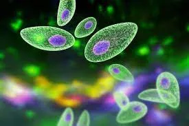
Toxoplasmosis is a parasitic infection caused by the Toxoplasma gondii parasite.Toxoplasma gondii is a parasitic organism classified as a member of the Coccidia class. Universally distributed.
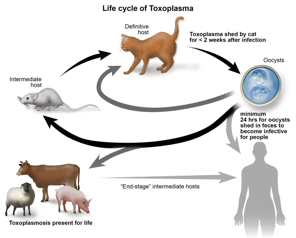
Transmission can occur through three distinct pathways: consumption of undercooked meat contaminated with cysts, exposure to oocysts found in cat feces, and transplacental transmission. Cats act as the primary host, while humans and other animals function as intermediate hosts.
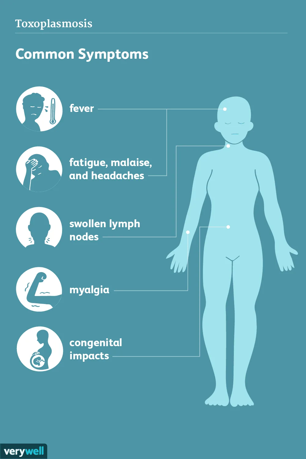
While infection is prevalent, the majority of persons do not show any symptoms. An acute illness has a resemblance to mononucleosis. Congenital infections can manifest as either asymptomatic or severe, presenting with symptoms such as encephalitis, hydrocephalus, chorioretinitis, brain calcifications, microcephalus, spontaneous miscarriage, and stillbirths. Immunocompromised patients may experience reactivation illness, which can manifest as encephalitis and multifocal lesions in the central nervous system.
Following infection by the cyst form, trophozoites proliferate and infiltrate the intestinal wall, where they are absorbed by macrophages. The trophozoites persist and proliferate within macrophages, circulate, and infiltrate various tissues, where they develop cysts that can remain alive for extended periods of time. Trophozoites spread through the placenta during the acute infection.
The diagnosis involves the use of serology, identification of trophozoites during the acute phase, and analysis of tissue biopsy specimens for cysts.
Typically, the combination of pyrimethamine and sulfadiazine is employed for the treatment of reactivation and congenital prevention disease. Antibodies have the ability to prevent the transmission of diseases from a mother to her fetus through the placenta. Pregnant women should refrain from handling cat litter boxes.
Prevention also encompasses the appropriate preparation of meat through thorough cooking.
Toxoplasmosis is a parasitic infection caused by the Toxoplasma gondii parasite.Toxoplasma gondii is a parasitic organism classified as a member of the Coccidia class. Universally distributed.
Transmission can occur through three distinct pathways: consumption of undercooked meat contaminated with cysts, exposure to oocysts found in cat feces, and transplacental transmission. Cats act as the primary host, while humans and other animals function as intermediate hosts.
While infection is prevalent, the majority of persons do not show any symptoms. An acute illness has a resemblance to mononucleosis. Congenital infections can manifest as either asymptomatic or severe, presenting with symptoms such as encephalitis, hydrocephalus, chorioretinitis, brain calcifications, microcephalus, spontaneous miscarriage, and stillbirths. Immunocompromised patients may experience reactivation illness, which can manifest as encephalitis and multifocal lesions in the central nervous system.
Following infection by the cyst form, trophozoites proliferate and infiltrate the intestinal wall, where they are absorbed by macrophages. The trophozoites persist and proliferate within macrophages, circulate, and infiltrate various tissues, where they develop cysts that can remain alive for extended periods of time. Trophozoites spread through the placenta during the acute infection.
The diagnosis involves the use of serology, identification of trophozoites during the acute phase, and analysis of tissue biopsy specimens for cysts.
Typically, the combination of pyrimethamine and sulfadiazine is employed for the treatment of reactivation and congenital prevention disease. Antibodies have the ability to prevent the transmission of diseases from a mother to her fetus through the placenta. Pregnant women should refrain from handling cat litter boxes.
Prevention also encompasses the appropriate preparation of meat through thorough cooking.
- Published on
Infectious Diseases and Microbiology - Pinworm Infection ( Enterobius vermicularis)
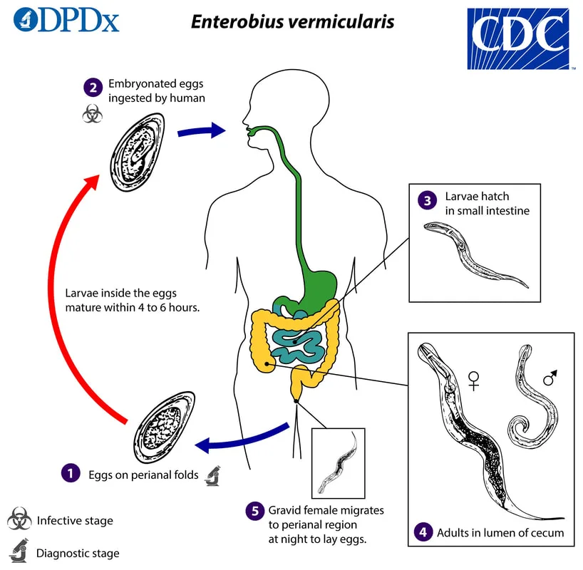
A roundworm known as Enterobius vermicularis inhabits the intestines.
The most prevalent nematode infection in the US is pinworm infection, which is caused by E vermicularis. Eggs can be consumed through fecal-oral spread or by eating dust that contains eggs. Fecal-oral autoinfection is a possible pathogen. When larvae grow in the perianal region and subsequently return through the rectum, retroinfection takes place.
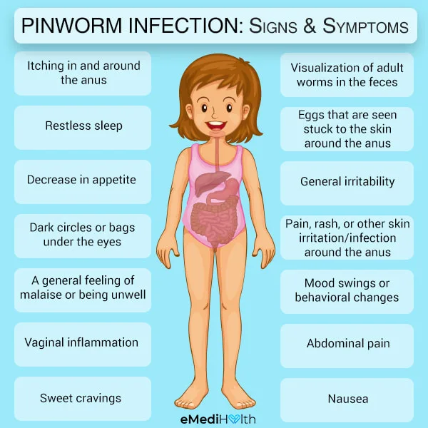
The main sign is pruritus, which can cause severe itching and subsequent bacterial infections.
An abnormal migration of the adult worm from the perineum might result in urethritis, vaginitis, salpingitis, or pelvic peritonitis.
After consumption, the small intestine is where eggs hatch, and the larvae move to the colon to undergo adult differentiation. During night, the mature gravid female leaves the colon and migrates outside the anus to lay eggs in the perianal region. A few hours later, the eggs become contagious.
By perianal sample with an applicator wrapped in transparent adhesive tape (sticky side out), eggs are collected for diagnosis. Stool samples typically don't contain eggs.
Mebendazole or pyrantel pamoate can be used to destroy mature worms. In order to stop reinfection, a second dose is administered during the two-week life cycle. Every family member ought to receive care.
Prevention entails timely medical attention and proper personal cleanliness.
A roundworm known as Enterobius vermicularis inhabits the intestines.
The most prevalent nematode infection in the US is pinworm infection, which is caused by E vermicularis. Eggs can be consumed through fecal-oral spread or by eating dust that contains eggs. Fecal-oral autoinfection is a possible pathogen. When larvae grow in the perianal region and subsequently return through the rectum, retroinfection takes place.
The main sign is pruritus, which can cause severe itching and subsequent bacterial infections.
An abnormal migration of the adult worm from the perineum might result in urethritis, vaginitis, salpingitis, or pelvic peritonitis.
After consumption, the small intestine is where eggs hatch, and the larvae move to the colon to undergo adult differentiation. During night, the mature gravid female leaves the colon and migrates outside the anus to lay eggs in the perianal region. A few hours later, the eggs become contagious.
By perianal sample with an applicator wrapped in transparent adhesive tape (sticky side out), eggs are collected for diagnosis. Stool samples typically don't contain eggs.
Mebendazole or pyrantel pamoate can be used to destroy mature worms. In order to stop reinfection, a second dose is administered during the two-week life cycle. Every family member ought to receive care.
Prevention entails timely medical attention and proper personal cleanliness.
- Published on
Infectious Diseases and Microbiology - Primary Meningoencephalitis Amebic
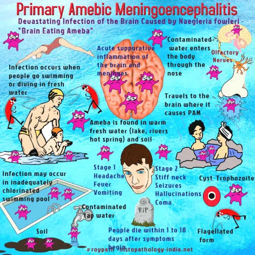
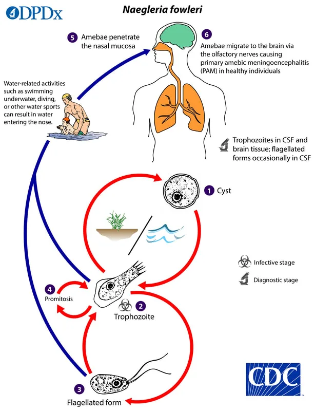
(Naegleria fowleri)
Physical: An opportunistic, free-living soil ameba.
By swimming in tainted warm fresh water, trophozoite infection is the mode of transmission to people.
An opportunistic, highly lethal meningoencephalitis caused by clinical N fowleri is marked by a fast-acting fever, stiff neck, headache, disorientation, vomiting, and changes in taste and smell perceptions.
Rapid progression results in a coma and death in 4-6 days.
Trophozoites penetrate the nasal mucosa and use the crebriform plate to access the central nervous system.
Cerebrospinal fluid examined under a microscope reveals neutrophils, erythrocytes, and trophozoites.
On a lawn of gram-negative rods, which are their food source, amebae can be cultivated.
Few people survive this sickness, which is extremely deadly. There has been some success with amphotericin B treatment in conjunction with rifampin and micoazole.
(Naegleria fowleri)
Physical: An opportunistic, free-living soil ameba.
By swimming in tainted warm fresh water, trophozoite infection is the mode of transmission to people.
An opportunistic, highly lethal meningoencephalitis caused by clinical N fowleri is marked by a fast-acting fever, stiff neck, headache, disorientation, vomiting, and changes in taste and smell perceptions.
Rapid progression results in a coma and death in 4-6 days.
Trophozoites penetrate the nasal mucosa and use the crebriform plate to access the central nervous system.
Cerebrospinal fluid examined under a microscope reveals neutrophils, erythrocytes, and trophozoites.
On a lawn of gram-negative rods, which are their food source, amebae can be cultivated.
Few people survive this sickness, which is extremely deadly. There has been some success with amphotericin B treatment in conjunction with rifampin and micoazole.
- Published on
Infectious Diseases and Miicrobiology - Giardiasis ( Giardia lamblia)
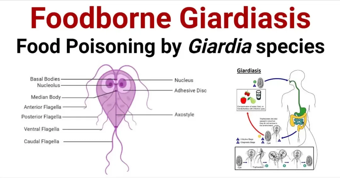
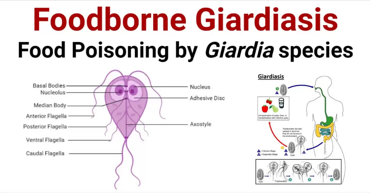
A flagellate protozoan that can take the shape of a cyst or trophozoite; the cyst is the infectious form.
Cyst ingestion is the mode of transmission, which can happen by oral-anal contact (homosexuals), fecal-oral dissemination (daycare facilities), or fecal pollution of water (community epidemiology epidemics). A typical source of infection for campers is mountain streams contaminated by animal waste from beavers, muskrats, people, and other animals.
Usually, 1-4 weeks after exposure, symptoms start to show. The illness might be asymptomatic, cause moderate watery diarrhea, or even worse, cause a malabsorption syndrome. Those without symptoms may develop into carriers and pass cysts in their stool. Abdominal cramps, bad-smelling feces, gas, and steatorrhea (fat in the stool) are the hallmarks of malabsorption syndrome, which can linger up to four weeks.
Trophozoites use their ventral sucking disk to cling to epithelial cells in the duodenum and jejunum. They do not penetrate, but they do prevent absorption—particularly of fat.
Four-nucleated cysts are observed in the produced stool of carriers who do not exhibit any symptoms. Diarrheal stool contains trophhozoites, which are pear-shaped bacteria with two nuclei, four flagella, and a ventral sucking disk. In addition to the string test, which is used to extract trophozoites from the duodenum in cases where stool tests yield negative results, there are several fecal antigen assays available.
Treatments such as metronidazole, furazolidone, or albendazole are frequently employed. Treatment with pomomycin is advised for pregnant patients who exhibit symptoms. In order to prevent infection, one must practice good hygiene, educate others about how infections spread, and take steps to filter, iodinate, or boil water from mountain streams.
A flagellate protozoan that can take the shape of a cyst or trophozoite; the cyst is the infectious form.
Cyst ingestion is the mode of transmission, which can happen by oral-anal contact (homosexuals), fecal-oral dissemination (daycare facilities), or fecal pollution of water (community epidemiology epidemics). A typical source of infection for campers is mountain streams contaminated by animal waste from beavers, muskrats, people, and other animals.
Usually, 1-4 weeks after exposure, symptoms start to show. The illness might be asymptomatic, cause moderate watery diarrhea, or even worse, cause a malabsorption syndrome. Those without symptoms may develop into carriers and pass cysts in their stool. Abdominal cramps, bad-smelling feces, gas, and steatorrhea (fat in the stool) are the hallmarks of malabsorption syndrome, which can linger up to four weeks.
Trophozoites use their ventral sucking disk to cling to epithelial cells in the duodenum and jejunum. They do not penetrate, but they do prevent absorption—particularly of fat.
Four-nucleated cysts are observed in the produced stool of carriers who do not exhibit any symptoms. Diarrheal stool contains trophhozoites, which are pear-shaped bacteria with two nuclei, four flagella, and a ventral sucking disk. In addition to the string test, which is used to extract trophozoites from the duodenum in cases where stool tests yield negative results, there are several fecal antigen assays available.
Treatments such as metronidazole, furazolidone, or albendazole are frequently employed. Treatment with pomomycin is advised for pregnant patients who exhibit symptoms. In order to prevent infection, one must practice good hygiene, educate others about how infections spread, and take steps to filter, iodinate, or boil water from mountain streams.

- Published on
Infectious Diseases and Microbiology - Trichomoniasis ( Trichomonas vaginalis)
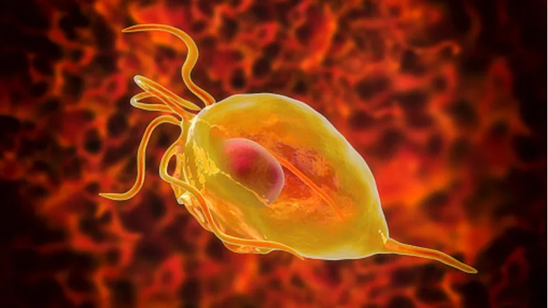
Trichomonas vaginalis is a type of protozoan that has flagella and can only be found in its active trophozoite form, without any cysts. It is marginally bigger than a granulocyte.
Principally spread by sexual intercourse. Commonly occurs alongside other sexually transmitted illnesses, such as gonorrhea. Can also be transmitted to newborn infants during the process of childbirth.
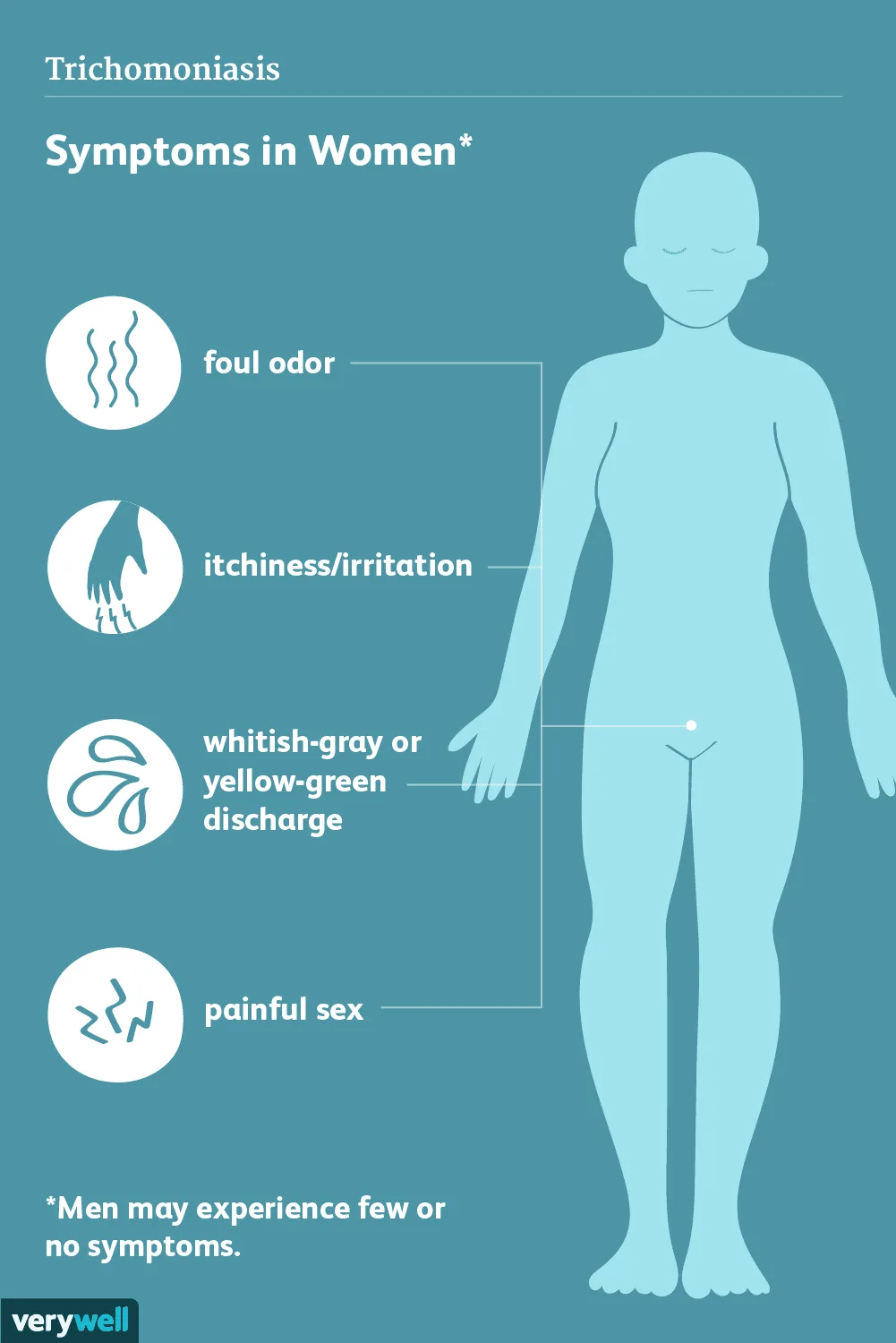
Often exhibiting symptoms. Women typically experience frothy vaginal discharge (vaginitis) as the primary symptom, along with minor vulvovaginal irritation. The discharge exhibits a greenish yellow hue and emits a musty aroma. Possible symptoms may consist of redness in the vagina and the presence of lesions on the cervix, sometimes referred to as "strawberry cervix". Typically, men do not show any symptoms, but they may experience urethritis, prostatitis, or epididymitis.
The presence of the trophozoite triggers an immune response in the host, leading to inflammation and damage to the epithelial cells.
Scientific testing facility The presence of oval trophozoites with four anterior flagella displaying erratic motions is a feature observed during the diagnosis by microscopic analysis of wet mounts. Additionally, antigen tests and culture tests are also accessible.
Both symptomatic women and their sexual partners can receive treatment with antibiotics, such as metronidazole, for prevention purposes. The most effective way to prevent is by adhering to safe sexual practices.
Trichomonas vaginalis is a type of protozoan that has flagella and can only be found in its active trophozoite form, without any cysts. It is marginally bigger than a granulocyte.
Principally spread by sexual intercourse. Commonly occurs alongside other sexually transmitted illnesses, such as gonorrhea. Can also be transmitted to newborn infants during the process of childbirth.
Often exhibiting symptoms. Women typically experience frothy vaginal discharge (vaginitis) as the primary symptom, along with minor vulvovaginal irritation. The discharge exhibits a greenish yellow hue and emits a musty aroma. Possible symptoms may consist of redness in the vagina and the presence of lesions on the cervix, sometimes referred to as "strawberry cervix". Typically, men do not show any symptoms, but they may experience urethritis, prostatitis, or epididymitis.
The presence of the trophozoite triggers an immune response in the host, leading to inflammation and damage to the epithelial cells.
Scientific testing facility The presence of oval trophozoites with four anterior flagella displaying erratic motions is a feature observed during the diagnosis by microscopic analysis of wet mounts. Additionally, antigen tests and culture tests are also accessible.
Both symptomatic women and their sexual partners can receive treatment with antibiotics, such as metronidazole, for prevention purposes. The most effective way to prevent is by adhering to safe sexual practices.
- Published on
Infectious Diseases and Microbiology - Balantidiasis ( Balantidium coli)
Balantidiasis is a medical condition.
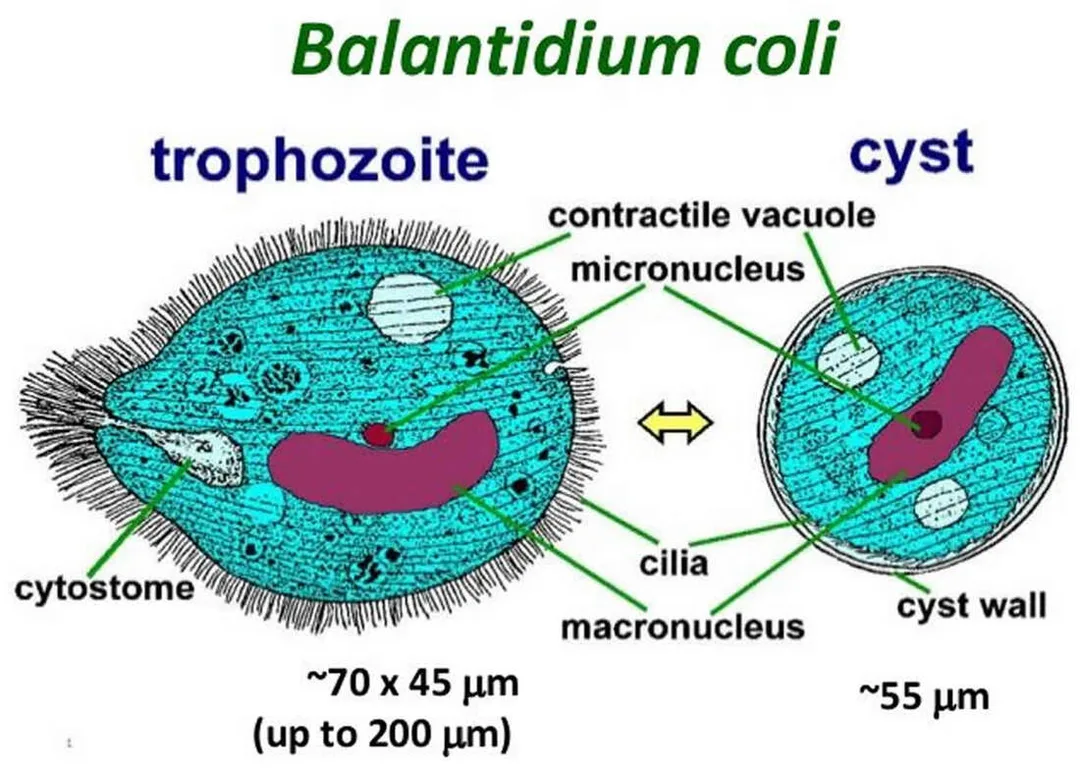
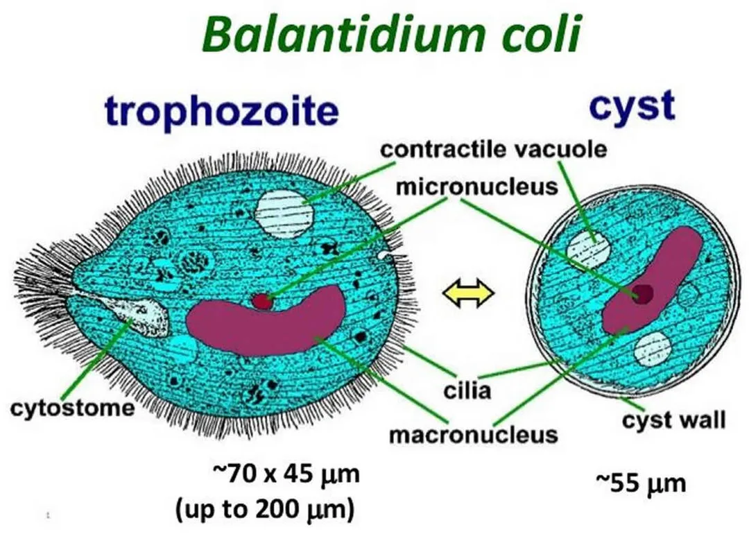
Balantidium coli is a single-celled parasite which causes balantidiasis.
An organism belonging to the group of protozoa that possess cilia. It is the largest disease-causing single-celled organism that is known to infect humans. Presence of cysts and trophozoite forms.
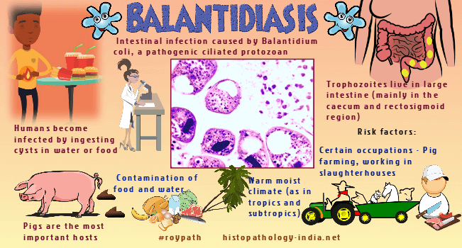
Transmission occurs when cyst-contaminated food or drink is ingested, or through the transmission of fecal matter via the oral route. Pigs are considered to be the main reservoir.
The majority of human infections are asymptomatic. This condition is an acute infection that presents with symptoms such as nausea, vomiting, stomach discomfort, and watery diarrhea that contains blood and mucus, resembling the symptoms observed in cases of E histolytica infection.
Upon ingestion, the cyst walls disintegrate, allowing the trophozoites to migrate to the colon where they nourish themselves by consuming bacteria and fecal matter. Infrequently, trophozoites infiltrate the mucosa and submucosa of the large bowel and terminal ileum, resulting in the formation of abscesses and ulcerations.
Cysts can be identified through a direct examination of produced stool. Trophozoites with cilia can be observed in fecal samples from individuals with diarrhea. The examination should be conducted swiftly as trophozoites rapidly deteriorate.
Tetracycline is the preferred medication. Iodoquinol and metronidazole serve as substitute medications.
Balantidiasis is a medical condition.
Balantidium coli is a single-celled parasite which causes balantidiasis.
An organism belonging to the group of protozoa that possess cilia. It is the largest disease-causing single-celled organism that is known to infect humans. Presence of cysts and trophozoite forms.
Transmission occurs when cyst-contaminated food or drink is ingested, or through the transmission of fecal matter via the oral route. Pigs are considered to be the main reservoir.
The majority of human infections are asymptomatic. This condition is an acute infection that presents with symptoms such as nausea, vomiting, stomach discomfort, and watery diarrhea that contains blood and mucus, resembling the symptoms observed in cases of E histolytica infection.
Upon ingestion, the cyst walls disintegrate, allowing the trophozoites to migrate to the colon where they nourish themselves by consuming bacteria and fecal matter. Infrequently, trophozoites infiltrate the mucosa and submucosa of the large bowel and terminal ileum, resulting in the formation of abscesses and ulcerations.
Cysts can be identified through a direct examination of produced stool. Trophozoites with cilia can be observed in fecal samples from individuals with diarrhea. The examination should be conducted swiftly as trophozoites rapidly deteriorate.
Tetracycline is the preferred medication. Iodoquinol and metronidazole serve as substitute medications.
- Published on
Infectious Diseases and Microbiology - Cryptosporidiosis (Cryptosporidium parvum)
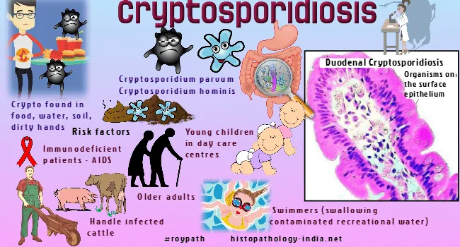
Cryptosporidiosis is an infectious disease caused by the parasite Cryptosporidium parvum.
Cryptosporidium parvum is a protozoan parasite that forms spores. Oocysts exhibit resistance to chlorination.
Transmission typically occurs when individuals consume water contaminated with oocysts or through direct contact with infected individuals via the fecal-oral or oral-anal routes. The duration of the incubation period ranges from 2 to 14 days.
Cryptosporidium parvum induces a transient, liquid bowel movement in persons with fully functional immune systems. Immunocompromised patients may experience chronic diarrhea, which can lead to significant fluid loss, malnutrition, electrolyte imbalance, and wasting.
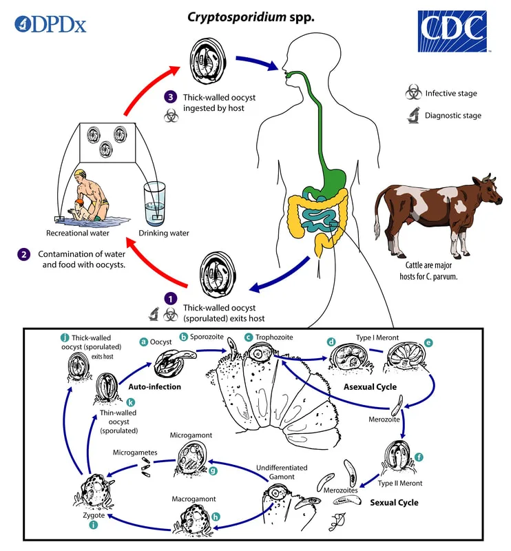
Following the consumption of oocysts, sporozoites mature and adhere to the epithelial cells of the jejunum, where asexual trophozoites emerge and multiply to generate merozoites. Merozoites undergo maturation into gametocytes in order to commence the process of sexual reproduction. The fertilized zygotes undergo development and transform into oocysts, which are ultimately expelled through feces.
The diagnosis entails doing fecal antigen tests and directly examining fecal smears for acid-fast oocysts using a microscope.
Immunocompromised persons have limited treatment options, with supportive measures being the primary approach.
Prophylaxis and rehydration. Paromomycin or azithromycin can potentially alleviate diarrhea. Individuals diagnosed with cryptosporidiosis should refrain from using recreational waters for a period of 2 weeks following the resolution of symptoms. The act of boiling water may be essential in order to prevent further instances in outbreaks caused by water contamination.
Cryptosporidiosis is an infectious disease caused by the parasite Cryptosporidium parvum.
Cryptosporidium parvum is a protozoan parasite that forms spores. Oocysts exhibit resistance to chlorination.
Transmission typically occurs when individuals consume water contaminated with oocysts or through direct contact with infected individuals via the fecal-oral or oral-anal routes. The duration of the incubation period ranges from 2 to 14 days.
Cryptosporidium parvum induces a transient, liquid bowel movement in persons with fully functional immune systems. Immunocompromised patients may experience chronic diarrhea, which can lead to significant fluid loss, malnutrition, electrolyte imbalance, and wasting.
Following the consumption of oocysts, sporozoites mature and adhere to the epithelial cells of the jejunum, where asexual trophozoites emerge and multiply to generate merozoites. Merozoites undergo maturation into gametocytes in order to commence the process of sexual reproduction. The fertilized zygotes undergo development and transform into oocysts, which are ultimately expelled through feces.
The diagnosis entails doing fecal antigen tests and directly examining fecal smears for acid-fast oocysts using a microscope.
Immunocompromised persons have limited treatment options, with supportive measures being the primary approach.
Prophylaxis and rehydration. Paromomycin or azithromycin can potentially alleviate diarrhea. Individuals diagnosed with cryptosporidiosis should refrain from using recreational waters for a period of 2 weeks following the resolution of symptoms. The act of boiling water may be essential in order to prevent further instances in outbreaks caused by water contamination.
- Published on
Infectious Diseases and Microbiology - Mucocutaneous Leishmaniasis (Leishmania species)
Mucocutaneous Leishmaniasis is a medical condition characterized by the presence of lesions affecting both the skin and mucous membranes.
Leishmania species
Several distinct species, such as L donovani, L tropica, L mexicana, and L braziliensis. This organism is an invasive flagellate that can be found in both blood and tissue.
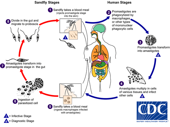
There are two distinct forms: the infective form with flagella, known as promastigote, and the nonflagellated form that lives inside cells, known as amastigote.
It is an obligate intracellular parasite that infects mononuclear phagocytes.
The sandfly serves as the vector for transmission. Several mammalian species can act as reservoirs.
Medical or pertaining to the practice of medicine. There are three primary clinical syndromes: Visceral leishmaniasis, also known as kala azar, is caused by the parasite Leishmania donovani. This parasite infiltrates macrophages, which are then transported to the liver, spleen, and bone marrow. The disease is characterized by a gradual onset of fever, chills, anemia, enlargement of the liver and spleen, and a decrease in the number of white blood cells. The disease duration is extended (spanning from months to years) and can lead to secondary infections, cachexia, and final mortality. Cutaneous leishmaniasis, also known as oriental sore or chiclero ulcers, is a skin infection caused by L tropica and L mexicana. It is characterized by the formation of a papule and subsequent ulceration at the site of infection.
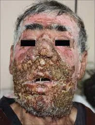
Mucocutaneous leishmaniasis, also known as espundia, is a condition caused by the parasite L braziliensis. It is characterized by the deformation of soft tissues, particularly in the nose and mouth, and frequently leads to other infections.
Amastigote forms are tested in lymph node aspirates, blood, spleen, liver, or bone marrow puncture (for L donovani), or in lymph node aspirates, scrapings, and biopsies from skin lesions (for L tropica, L mexicana, and L braziliensis). Leishmanin skin tests yield good results upon recovery, but display negative results during the presence of ongoing disease.
There are additional serologic testing available.
Sodium stibogluconate, a kind of pentavalent antimonial, is employed for therapeutic purposes. Prevention entails the utilization of insect repellents, reducing outside activities between the hours of twilight to morning, and employing physical barriers such as fine-mesh bed netting and protective gear to avoid being bitten by sandflies.
Mucocutaneous Leishmaniasis is a medical condition characterized by the presence of lesions affecting both the skin and mucous membranes.
Leishmania species
Several distinct species, such as L donovani, L tropica, L mexicana, and L braziliensis. This organism is an invasive flagellate that can be found in both blood and tissue.
There are two distinct forms: the infective form with flagella, known as promastigote, and the nonflagellated form that lives inside cells, known as amastigote.
It is an obligate intracellular parasite that infects mononuclear phagocytes.
The sandfly serves as the vector for transmission. Several mammalian species can act as reservoirs.
Medical or pertaining to the practice of medicine. There are three primary clinical syndromes: Visceral leishmaniasis, also known as kala azar, is caused by the parasite Leishmania donovani. This parasite infiltrates macrophages, which are then transported to the liver, spleen, and bone marrow. The disease is characterized by a gradual onset of fever, chills, anemia, enlargement of the liver and spleen, and a decrease in the number of white blood cells. The disease duration is extended (spanning from months to years) and can lead to secondary infections, cachexia, and final mortality. Cutaneous leishmaniasis, also known as oriental sore or chiclero ulcers, is a skin infection caused by L tropica and L mexicana. It is characterized by the formation of a papule and subsequent ulceration at the site of infection.
Mucocutaneous leishmaniasis, also known as espundia, is a condition caused by the parasite L braziliensis. It is characterized by the deformation of soft tissues, particularly in the nose and mouth, and frequently leads to other infections.
Amastigote forms are tested in lymph node aspirates, blood, spleen, liver, or bone marrow puncture (for L donovani), or in lymph node aspirates, scrapings, and biopsies from skin lesions (for L tropica, L mexicana, and L braziliensis). Leishmanin skin tests yield good results upon recovery, but display negative results during the presence of ongoing disease.
There are additional serologic testing available.
Sodium stibogluconate, a kind of pentavalent antimonial, is employed for therapeutic purposes. Prevention entails the utilization of insect repellents, reducing outside activities between the hours of twilight to morning, and employing physical barriers such as fine-mesh bed netting and protective gear to avoid being bitten by sandflies.