- Published on
Dermatology - Merkel Cell Carcinoma
Merkel cell carcinoma, also known as cutaneous neuroendocrine tumor, is an uncommon form of solid tumor that is mostly found in Caucasians and often manifests beyond the age of 50. Individuals with immunosuppression experience it 10–30 times more frequently.
The tumor can appear on the head or the extremities, and it can be single or numerous.
Damage
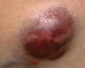
The lesion is a pink, red to violet, or reddish brown, dome-shaped, cutaneous to subcutaneous papule, nodule, or tumor (0.5–5 cm), generally solitary. Greater lesions have the potential to ulcerate, although the skin above remains intact.
The diagnosis is made clinically, and immunohistopathology and biopsy confirm it.
Excision and Mohs surgical repair are the methods of treatment. Given the high incidence of regional metastases, proactive regional node dissection or sentinel node biopsy is recommended.
With the exception of extremely tiny lesions, radiation therapy is usually necessary. The prognosis is uncertain and recurrence rates are high even with treatment.
Merkel cell carcinoma, also known as cutaneous neuroendocrine tumor, is an uncommon form of solid tumor that is mostly found in Caucasians and often manifests beyond the age of 50. Individuals with immunosuppression experience it 10–30 times more frequently.
The tumor can appear on the head or the extremities, and it can be single or numerous.
Damage
The lesion is a pink, red to violet, or reddish brown, dome-shaped, cutaneous to subcutaneous papule, nodule, or tumor (0.5–5 cm), generally solitary. Greater lesions have the potential to ulcerate, although the skin above remains intact.
The diagnosis is made clinically, and immunohistopathology and biopsy confirm it.
Excision and Mohs surgical repair are the methods of treatment. Given the high incidence of regional metastases, proactive regional node dissection or sentinel node biopsy is recommended.
With the exception of extremely tiny lesions, radiation therapy is usually necessary. The prognosis is uncertain and recurrence rates are high even with treatment.
- Published on
Dermatology - Fixed Drug Reaction
An unfavorable cutaneous reaction to an ingested medicine is known as a fixed drug eruption, and it is typified by the development of a single (or sometimes several) erythematous patch or plaque. Tetracyclines, sulfa medications, metronidazole, nystatin, salicylates, NSAIDs, phenylbutazone, phenacetin, barbiturates, oral contraceptives, quinine, quinidine, phenolphthalein, and food coloring (yellow) are the most often implicated substances. Within hours of consumption, if the patient is reexposed to the offending drug, the FDE reoccurs at the same (fixed) skin spot.
In a previously sensitized person, lesions might develop 30 minutes to 8 hours after drug administration and can be itchy, unpleasant, or burning. Lesions go away in a few days to a few weeks after the medication is stopped.
Damage
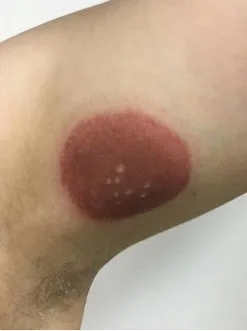
Hours after ingesting the offending medicine, a well-defined patch of erythema turns into a dusky red to violaceous macule or plaque. Lesions are usually solitary and can grow to be rather large, although they can also be many and dispersed randomly.
Lesions may progress to painful bullae and eventually erosions. Postinflammatory hyperpigmentation with a dark brown to violet tint may appear after recovery. Although the genital area is most commonly affected, other sites such as the perioral, periorbital, conjunctival, and oropharyngeal regions may also be affected.
Once the offending medication is stopped, the lesions must heal for the diagnosis to be considered clinical.
Give up the problematic substance. Apply antibacterial ointment to erosions. Oral prednisone at a dose of 1 mg/kg body weight decreased over a period of 2 weeks is recommended for mucosal lesions that are extensive, generalized, and extremely painful.
An unfavorable cutaneous reaction to an ingested medicine is known as a fixed drug eruption, and it is typified by the development of a single (or sometimes several) erythematous patch or plaque. Tetracyclines, sulfa medications, metronidazole, nystatin, salicylates, NSAIDs, phenylbutazone, phenacetin, barbiturates, oral contraceptives, quinine, quinidine, phenolphthalein, and food coloring (yellow) are the most often implicated substances. Within hours of consumption, if the patient is reexposed to the offending drug, the FDE reoccurs at the same (fixed) skin spot.
In a previously sensitized person, lesions might develop 30 minutes to 8 hours after drug administration and can be itchy, unpleasant, or burning. Lesions go away in a few days to a few weeks after the medication is stopped.
Damage
Hours after ingesting the offending medicine, a well-defined patch of erythema turns into a dusky red to violaceous macule or plaque. Lesions are usually solitary and can grow to be rather large, although they can also be many and dispersed randomly.
Lesions may progress to painful bullae and eventually erosions. Postinflammatory hyperpigmentation with a dark brown to violet tint may appear after recovery. Although the genital area is most commonly affected, other sites such as the perioral, periorbital, conjunctival, and oropharyngeal regions may also be affected.
Once the offending medication is stopped, the lesions must heal for the diagnosis to be considered clinical.
Give up the problematic substance. Apply antibacterial ointment to erosions. Oral prednisone at a dose of 1 mg/kg body weight decreased over a period of 2 weeks is recommended for mucosal lesions that are extensive, generalized, and extremely painful.
- Published on
Dermatology - Darier Disease
Due to a loss-of-function mutation in the ATP2A2 gene, Darier disease is an uncommon, autosomal-dominant, hereditary condition with a late start and symptoms related to the mucocutaneous and nail areas.
Painful, itchy, and malodorous lesions are present. They are often made worse by the summer's heat and humidity, as well as by UVB rays, physical damage, and bacterial infections. Less commonly than diminished intelligence, the illness is linked to affective disorders.
Damage
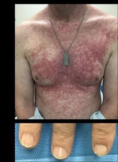
There are numerous, distinct, itchy papules that are crusted and scaling. An hole that resembles a slit is revealed upon removal of the scaling crust. Papules congregate to create enormous plaques, especially in intertriginous areas, which are covered in hypertrophic warty masses that smell bad. Although the hair are unaffected, severe scalp involvement and scarring may lead to irreversible alopecia. The nails have a distinctive V-shaped scalloping, are thin, and split distally. Mucocutaneous lesions, also referred to as "cobblestone" lesions, are white, centrally depressed papules on the mucosa of the cheeks, palate, and gums.
The history of familial involvement, clinical presentation, and histology all play a role in the diagnosis.
The differential diagnosis consists of Grover disease, pemphigus foliaceus, benign familial pemphigus (also known as Hayley-Hailey disease), seborrheic dermatitis, and flat warts (verrucae planae).
Advise patients to refrain from rubbing and friction, and to wear sunscreen. To inhibit bacterial infection, administer systemic and topical antibiotic therapy. Retinoids, either systemic (acitretin or isotretinoin) or topical (tazarotene and adjapalene), provide additional advantages.
Due to a loss-of-function mutation in the ATP2A2 gene, Darier disease is an uncommon, autosomal-dominant, hereditary condition with a late start and symptoms related to the mucocutaneous and nail areas.
Painful, itchy, and malodorous lesions are present. They are often made worse by the summer's heat and humidity, as well as by UVB rays, physical damage, and bacterial infections. Less commonly than diminished intelligence, the illness is linked to affective disorders.
Damage
There are numerous, distinct, itchy papules that are crusted and scaling. An hole that resembles a slit is revealed upon removal of the scaling crust. Papules congregate to create enormous plaques, especially in intertriginous areas, which are covered in hypertrophic warty masses that smell bad. Although the hair are unaffected, severe scalp involvement and scarring may lead to irreversible alopecia. The nails have a distinctive V-shaped scalloping, are thin, and split distally. Mucocutaneous lesions, also referred to as "cobblestone" lesions, are white, centrally depressed papules on the mucosa of the cheeks, palate, and gums.
The history of familial involvement, clinical presentation, and histology all play a role in the diagnosis.
The differential diagnosis consists of Grover disease, pemphigus foliaceus, benign familial pemphigus (also known as Hayley-Hailey disease), seborrheic dermatitis, and flat warts (verrucae planae).
Advise patients to refrain from rubbing and friction, and to wear sunscreen. To inhibit bacterial infection, administer systemic and topical antibiotic therapy. Retinoids, either systemic (acitretin or isotretinoin) or topical (tazarotene and adjapalene), provide additional advantages.
- Published on
Dermatology - Pattern Hair Loss
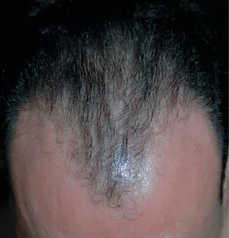
The most prevalent kind of progressive baldness, pattern hair loss is brought on by an androgen's activity and genetic predisposition. Men may experience bitemporal recession, frontal and/or vertex thinning, or complete hair loss. The pattern can start at any point after puberty and may fully manifest in the second decade of life. It happens later in life, typically in the sixth decade, in women, where it is far less common.
The majority of individuals arrive with baldness or gradually losing hair. Due to the importance that our society placed on having a "healthy head of hair," many people find the cosmetic look of pattern hair loss to be extremely upsetting. Signs of androgen excess in young women include virilization, irregular menses, acne, and hirsutism. These should be looked for as serious issues. The majority of women who experience pattern hair loss, however, have normal endocrinology.
Skin findings are rare; scalps with advanced pattern hair loss are glossy and smooth, and follicle orifices are hardly noticeable to the untrained eye. In pattern hair loss areas, the texture of the hair becomes finer (shorter in length, reduced diameter). Hair eventually atrophies totally and turns vellus.
Clinical diagnosis is occasionally confirmed by biopsy. Diffuse nonscarring alopecia, trichotillomania, iron deficiency, hypothyroidism, hyperthyroidism, secondary syphilis, telogen effluvium, and seborrheic dermatitis are among the differential diagnoses.
Administering a daily dosage of 1 mg of oral finasteride can potentially decelerate hair loss within a span of three months. After six months, there is a possibility of regeneration of fully developed hair on the top and front areas of the scalp. It is crucial to maintain the usage of the medicine, as discontinuation may reverse the effects. However, it is important to note that there is a potential danger of experiencing a drop in sexual desire and difficulties in achieving or maintaining an erection.
Minoxidil 2% and 5% solutions administered topically may help slow down the pace of hair loss or partially regrow hair that has been lost. Spironolactone, cyproterone acetate, flutamide, and cimetidine are beneficial in women with increased adrenal androgens; however, they should not be administered in men. Other possibilities include hairpieces and hair replacement surgery.
The most prevalent kind of progressive baldness, pattern hair loss is brought on by an androgen's activity and genetic predisposition. Men may experience bitemporal recession, frontal and/or vertex thinning, or complete hair loss. The pattern can start at any point after puberty and may fully manifest in the second decade of life. It happens later in life, typically in the sixth decade, in women, where it is far less common.
The majority of individuals arrive with baldness or gradually losing hair. Due to the importance that our society placed on having a "healthy head of hair," many people find the cosmetic look of pattern hair loss to be extremely upsetting. Signs of androgen excess in young women include virilization, irregular menses, acne, and hirsutism. These should be looked for as serious issues. The majority of women who experience pattern hair loss, however, have normal endocrinology.
Skin findings are rare; scalps with advanced pattern hair loss are glossy and smooth, and follicle orifices are hardly noticeable to the untrained eye. In pattern hair loss areas, the texture of the hair becomes finer (shorter in length, reduced diameter). Hair eventually atrophies totally and turns vellus.
Clinical diagnosis is occasionally confirmed by biopsy. Diffuse nonscarring alopecia, trichotillomania, iron deficiency, hypothyroidism, hyperthyroidism, secondary syphilis, telogen effluvium, and seborrheic dermatitis are among the differential diagnoses.
Administering a daily dosage of 1 mg of oral finasteride can potentially decelerate hair loss within a span of three months. After six months, there is a possibility of regeneration of fully developed hair on the top and front areas of the scalp. It is crucial to maintain the usage of the medicine, as discontinuation may reverse the effects. However, it is important to note that there is a potential danger of experiencing a drop in sexual desire and difficulties in achieving or maintaining an erection.
Minoxidil 2% and 5% solutions administered topically may help slow down the pace of hair loss or partially regrow hair that has been lost. Spironolactone, cyproterone acetate, flutamide, and cimetidine are beneficial in women with increased adrenal androgens; however, they should not be administered in men. Other possibilities include hairpieces and hair replacement surgery.
- Published on
Dermatology-Pearly Penile Papules
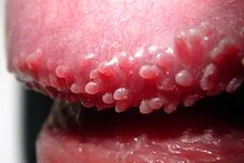
A typical variation in anatomy, pearly penile papules affect up to 19% of men.Pearly penile papules are asymptomatic, though initially they may cause some concern. They are skin-colored, distinct, domed papules that range in size from 1 to 2 mm and are equally spaced throughout the corona, resembling cobblestones.
The differential includes molluscum contagiosum and condylomata acuminatum. The diagnosis is clinical.
The confirmation that these are typical anatomic features is all that is needed for management.
A typical variation in anatomy, pearly penile papules affect up to 19% of men.Pearly penile papules are asymptomatic, though initially they may cause some concern. They are skin-colored, distinct, domed papules that range in size from 1 to 2 mm and are equally spaced throughout the corona, resembling cobblestones.
The differential includes molluscum contagiosum and condylomata acuminatum. The diagnosis is clinical.
The confirmation that these are typical anatomic features is all that is needed for management.
- Published on
Dermatology - Epidermal Inclusion Cyst
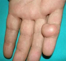
Traumatic epidermis implantation into the dermis results in an epidermal inclusion cyst. The dermis is where traumatized grafted epidermis grows, and the cyst cavity contains keratin buildup that is surrounded by a stratified squamous epithelium with a well-formed granular layer.
The lesion typically affects the palms, soles, and fingers and manifests as a cutaneous nodule.The more superficial nature of pidermal inclusion cysts (found in the dermis rather than the subcutaneous fat) and their overlaying punctum set them apart from lipomas. Although they may appear similar on the surface to epidermal inclusion cysts, many different benign and malignant tumors do not have the punctutum.
If there are no symptoms, treatment is not necessary. Incision and drainage or intralesional triamcinolone acetomide (5–10 mg/mL) are two possible treatments for inflamed lesions. Surgical removal of big or symptomatic cysts is therapeutic.
Traumatic epidermis implantation into the dermis results in an epidermal inclusion cyst. The dermis is where traumatized grafted epidermis grows, and the cyst cavity contains keratin buildup that is surrounded by a stratified squamous epithelium with a well-formed granular layer.
The lesion typically affects the palms, soles, and fingers and manifests as a cutaneous nodule.The more superficial nature of pidermal inclusion cysts (found in the dermis rather than the subcutaneous fat) and their overlaying punctum set them apart from lipomas. Although they may appear similar on the surface to epidermal inclusion cysts, many different benign and malignant tumors do not have the punctutum.
If there are no symptoms, treatment is not necessary. Incision and drainage or intralesional triamcinolone acetomide (5–10 mg/mL) are two possible treatments for inflamed lesions. Surgical removal of big or symptomatic cysts is therapeutic.
- Published on
nDermatology - Agiosarcoma
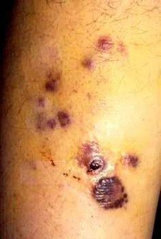
An very unusual and malignant growth of endothelial cells is called angiosarcoma.
Purpuric macules, papules, brilliant red or violaceous nodules, and even black or solid nodules that bleed readily and ulcerate are examples of lesions. Normal skin typically has lesions on the scalp and upper forehead, or there may be localized lymphedema, such as post-irradiation lymphedema or Stuart-Treves syndrome after a mastectomy.
Clinical diagnosis is made, and a biopsy confirms the diagnosis. Other vascular lesions, lymphoma, pyogenic granuloma, and Kaposi sarcoma are among the differential diagnoses.
Treatment consists of wide-margin excision and chemotherapy (paclitaxel or liposomal doxorubicin); nonetheless, the 5-year survival rate is still somewhat higher than 10% even with treatment.
An very unusual and malignant growth of endothelial cells is called angiosarcoma.
Purpuric macules, papules, brilliant red or violaceous nodules, and even black or solid nodules that bleed readily and ulcerate are examples of lesions. Normal skin typically has lesions on the scalp and upper forehead, or there may be localized lymphedema, such as post-irradiation lymphedema or Stuart-Treves syndrome after a mastectomy.
Clinical diagnosis is made, and a biopsy confirms the diagnosis. Other vascular lesions, lymphoma, pyogenic granuloma, and Kaposi sarcoma are among the differential diagnoses.
Treatment consists of wide-margin excision and chemotherapy (paclitaxel or liposomal doxorubicin); nonetheless, the 5-year survival rate is still somewhat higher than 10% even with treatment.
- Published on
Dermatology - Scabies
The mite Sarcoptes scabiei var. hominis causes scabies, an epidermal infestation that is spread by skin-to-skin contact and bites. On clothes or bedding, mites can survive for more than two days.
Patients frequently report experiencing such symptoms in close relatives or romantic partners.
Intense, pervasive pruritus typically spares the head and neck and disrupts sleep. Atopy-prone individuals may develop eczematous dermatitis, but other individuals may have pruritus for several months without any visible symptoms. There is a chance of secondary infection, which manifests as tenderness.
Damage
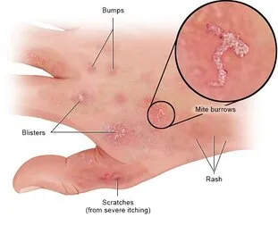
Hypersensitivity, lesions brought on by persistent rubbing and scratching, and secondary infection are among the lesions that appear at the infestation site. Skin-colored, linear or serpiginous ridges, 0.5–1 cm long, with a tiny vesicle or papule at the end of the tunnel, are present at the infection site. Burrows typically occur in regions with few or no hair follicles, typically in the thin and delicate stratum corneum (the interdigital webs of hands, wrists, penis shafts, elbows, feet, buttocks, and axillae). Burrows are 5 mm on average, although they can reach up to 10 cm in length.
Nodules that range in size from 5 to 20 mm and are smooth, red, pink, tan, or brown may appear.
Widespread, tiny, urticarial, edematous papules, mainly on the anterior trunk, thighs, buttocks, and forearms, are indicative of hypersensitivity reactions. There may be hypo- and hyperpigmentation following an inflammation.
Clinical diagnosis and, if feasible, microscopy (detection of mites, eggs, or mite excrement) are used to confirm the diagnosis. Atopic dermatitis, allergic contact dermatitis, metabolic pruritus, urticaria pigmentosa, papular urticaria, prurigo nodularis, and pseudolymphoma are among the conditions on the differential.
Treat infected people concurrently with those in close physical contact, regardless of the presence of symptoms. Apply lindane (g-benzenehexachloride) 1% lotion or cream to every part of the body starting at the neck and washing it off completely after 8 hours, or permethrin 5% cream from head to toe. Lindane shouldn't be used by patients with significant dermatitis, women who are pregnant or nursing, children under the age of two, or after taking a bath or shower. There is lindane resistance in mites. In many nations, lindane is a crucial substitute due to its low cost. As an alternative, apply 10% crotamiton, 2%–10% sulfur in petrolatum, 10% and 25% benzyl benzoate, benzyl benzoate with sulfiram, 0.5% malathion, 25% sulfram, and 200 μg/kg of ivermectin. Intralesional triamcinolone, 5–10 mg/mL into each lesion, works well for scabietic nodules.
The mite Sarcoptes scabiei var. hominis causes scabies, an epidermal infestation that is spread by skin-to-skin contact and bites. On clothes or bedding, mites can survive for more than two days.
Patients frequently report experiencing such symptoms in close relatives or romantic partners.
Intense, pervasive pruritus typically spares the head and neck and disrupts sleep. Atopy-prone individuals may develop eczematous dermatitis, but other individuals may have pruritus for several months without any visible symptoms. There is a chance of secondary infection, which manifests as tenderness.
Damage
Hypersensitivity, lesions brought on by persistent rubbing and scratching, and secondary infection are among the lesions that appear at the infestation site. Skin-colored, linear or serpiginous ridges, 0.5–1 cm long, with a tiny vesicle or papule at the end of the tunnel, are present at the infection site. Burrows typically occur in regions with few or no hair follicles, typically in the thin and delicate stratum corneum (the interdigital webs of hands, wrists, penis shafts, elbows, feet, buttocks, and axillae). Burrows are 5 mm on average, although they can reach up to 10 cm in length.
Nodules that range in size from 5 to 20 mm and are smooth, red, pink, tan, or brown may appear.
Widespread, tiny, urticarial, edematous papules, mainly on the anterior trunk, thighs, buttocks, and forearms, are indicative of hypersensitivity reactions. There may be hypo- and hyperpigmentation following an inflammation.
Clinical diagnosis and, if feasible, microscopy (detection of mites, eggs, or mite excrement) are used to confirm the diagnosis. Atopic dermatitis, allergic contact dermatitis, metabolic pruritus, urticaria pigmentosa, papular urticaria, prurigo nodularis, and pseudolymphoma are among the conditions on the differential.
Treat infected people concurrently with those in close physical contact, regardless of the presence of symptoms. Apply lindane (g-benzenehexachloride) 1% lotion or cream to every part of the body starting at the neck and washing it off completely after 8 hours, or permethrin 5% cream from head to toe. Lindane shouldn't be used by patients with significant dermatitis, women who are pregnant or nursing, children under the age of two, or after taking a bath or shower. There is lindane resistance in mites. In many nations, lindane is a crucial substitute due to its low cost. As an alternative, apply 10% crotamiton, 2%–10% sulfur in petrolatum, 10% and 25% benzyl benzoate, benzyl benzoate with sulfiram, 0.5% malathion, 25% sulfram, and 200 μg/kg of ivermectin. Intralesional triamcinolone, 5–10 mg/mL into each lesion, works well for scabietic nodules.
- Published on
Dermatology - Hand-Foot-and-Mouth Disease
The most frequent enteroviruses that cause hand, foot, and mouth illness are enterovirus 71 and coxsackie virus A16. In temperate climes, the fall and early summer months are when children under the age of ten are most typically afflicted. The virus is extremely contagious and spreads from person to person through the oral and fecal-oral pathways.
Children's painful oral mucosal sores often cause them to shun food. There may also be other moderate constitutional symptoms such as diarrhea, acute malaise, high fever, and joint pain. Lung involvement and CNS (aseptic meningitis, encephalitis, meningoencephalitis, flaccid paralysis) may be linked to EV17 infections.
Damage
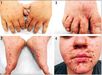
Usually, 5–10 unpleasant, grayish vesicles in the oral mucosa ulcerate and cause pain. On the distal extremities, cutaneous lesions that may be painful and sensitive or asymptomatic develop soon after or simultaneously. The macules and papules that swiftly develop into vesicles make up the cutaneous exanthem, which is typically found on the palms and soles, particularly on the sides of the fingers, toes, and buttocks. These vesicles typically don't burst and may have a distinctive "linear" shape. Ruptures of vesicles can cause crusts and erosions at other cutaneous locations. Wounds resolve without leaving scars.
The diagnosis of a sudden breakout of lesions in the distal extremities and mouth is usually clinical, however the virus can also be identified from stool samples, vesicles, and throat washes. The differential includes HSV infection, aphthous stomatitis, herpangina, erythema multiforme, and adverse medication reaction if the oral lesions are the only ones seen.
Treatment for the virus is symptomatic and supportive since it is self-limiting.
The most frequent enteroviruses that cause hand, foot, and mouth illness are enterovirus 71 and coxsackie virus A16. In temperate climes, the fall and early summer months are when children under the age of ten are most typically afflicted. The virus is extremely contagious and spreads from person to person through the oral and fecal-oral pathways.
Children's painful oral mucosal sores often cause them to shun food. There may also be other moderate constitutional symptoms such as diarrhea, acute malaise, high fever, and joint pain. Lung involvement and CNS (aseptic meningitis, encephalitis, meningoencephalitis, flaccid paralysis) may be linked to EV17 infections.
Damage
Usually, 5–10 unpleasant, grayish vesicles in the oral mucosa ulcerate and cause pain. On the distal extremities, cutaneous lesions that may be painful and sensitive or asymptomatic develop soon after or simultaneously. The macules and papules that swiftly develop into vesicles make up the cutaneous exanthem, which is typically found on the palms and soles, particularly on the sides of the fingers, toes, and buttocks. These vesicles typically don't burst and may have a distinctive "linear" shape. Ruptures of vesicles can cause crusts and erosions at other cutaneous locations. Wounds resolve without leaving scars.
The diagnosis of a sudden breakout of lesions in the distal extremities and mouth is usually clinical, however the virus can also be identified from stool samples, vesicles, and throat washes. The differential includes HSV infection, aphthous stomatitis, herpangina, erythema multiforme, and adverse medication reaction if the oral lesions are the only ones seen.
Treatment for the virus is symptomatic and supportive since it is self-limiting.
- Published on
Dermatology - Solar Lentigo
Solar Lentigo
Localized melanocyte proliferation brought on by either acute or long-term UV exposure is the cause of solar lentigines. The onset typically occurs beyond the age of 40, with lighter-skinned people having a higher incidence.
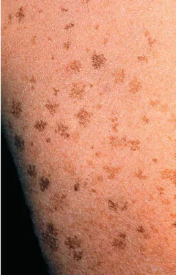
One to three centimeter and up to five centimeter macules can be pale yellow, light brown, dark brown, or a variegated mix of brown. Lesions are ill-defined, round, and have slightly uneven boundaries. After an acute sunburn, lesions are generally the same size, stellate, strongly defined, and dispersed and discrete.
The differential includes freckles, seborrheic keratosis, spreading pigmented actinic keratosis, and lentigo maligna. The diagnosis is clinical.
Treatment for benign solar lentigines is not necessary; however, if removal is desired, cryosurgery or laser surgery are feasible options.
Solar Lentigo
Localized melanocyte proliferation brought on by either acute or long-term UV exposure is the cause of solar lentigines. The onset typically occurs beyond the age of 40, with lighter-skinned people having a higher incidence.
One to three centimeter and up to five centimeter macules can be pale yellow, light brown, dark brown, or a variegated mix of brown. Lesions are ill-defined, round, and have slightly uneven boundaries. After an acute sunburn, lesions are generally the same size, stellate, strongly defined, and dispersed and discrete.
The differential includes freckles, seborrheic keratosis, spreading pigmented actinic keratosis, and lentigo maligna. The diagnosis is clinical.
Treatment for benign solar lentigines is not necessary; however, if removal is desired, cryosurgery or laser surgery are feasible options.