- Published on
Dermatology - Phimosis
Phimosis
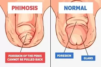
Lichen sclerosus, lichen planus, cicatricial pemphigoid, chronic lymphedema, Kaposi sarcoma, and nonspecific balanoposthitis (inflammation of the foreskin or prepuce) can all result in phimosis, an irretractable foreskin. It is not possible to check the glans for precancerous alterations when phimosis is present.
The terminal stage of chronic phimosis, known as balanitis xerotica obliterans, is characterized by a fibrotic, contracted foreskin that is fastened over glans and cannot be retracted.
The foreskin may enlarge, there may be pain, edema, or trouble peeing. The glans penis tip does not allow the foreskin to be removed.
Clinical diagnosis is made, and the goal of the differential is to find any underlying illnesses.
Attend to any underlying illness. Phimosis can be treated by surgically separating or removing the foreskin, or by gently expanding it gradually.
Phimosis
Lichen sclerosus, lichen planus, cicatricial pemphigoid, chronic lymphedema, Kaposi sarcoma, and nonspecific balanoposthitis (inflammation of the foreskin or prepuce) can all result in phimosis, an irretractable foreskin. It is not possible to check the glans for precancerous alterations when phimosis is present.
The terminal stage of chronic phimosis, known as balanitis xerotica obliterans, is characterized by a fibrotic, contracted foreskin that is fastened over glans and cannot be retracted.
The foreskin may enlarge, there may be pain, edema, or trouble peeing. The glans penis tip does not allow the foreskin to be removed.
Clinical diagnosis is made, and the goal of the differential is to find any underlying illnesses.
Attend to any underlying illness. Phimosis can be treated by surgically separating or removing the foreskin, or by gently expanding it gradually.
- Published on
Dermatology - Plaque Psoriasis
Psoriasis is usually a chronic condition. Environmental triggers include stress, bacterial infections, trauma, and certain medicines, in addition to polygenic predisposition. Any age can be the onset, with ages 8, 22, and 55 being the peaks. More severe disease is predicted by an early beginning. Lesions that persist for months or years affect the majority of patients.
Pruritus is prevalent, particularly in the anogenital and scalp areas.
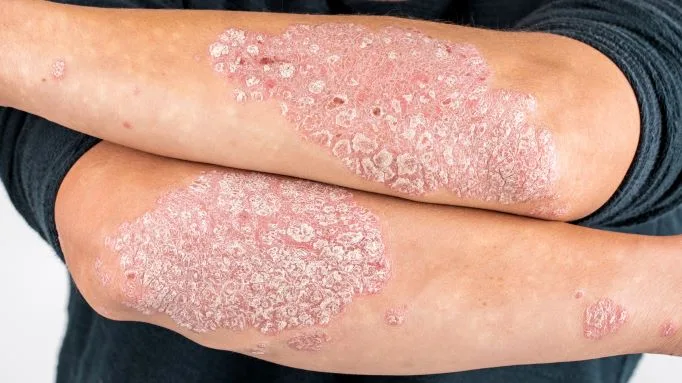
Polycyclic, geographic lesions can grow from sharply marginated, dull-red plaques with loose lamellar, silvery-white scales. These could exhibit arciform, serpiginous, and annular patterns if they partially retreat. Unless the lesion is very persistent, lamellar scales can be easily eliminated. The sacralgluteal area, scalp, elbows, knees, palms, and soles are the preference sites for localizing lesions, which can be single or many. Lesions frequently spare exposed areas and exhibit bilateral symmetry. Massive hyperkeratosis that is glossy white or yellowish on the palms and soles, with a clearly defined base, is present. It is difficult to eradicate. Bleeding, painful fissures, and cracking are possible.
Plaques in the body folds are macerated, frequently bright red and fissured (inverse psoriasis), and lesions from intertrigo, candidiasis, or contact dermatitis can only be distinguished by a strong demarcation. Pitting, subungual hyperkeratosis, onycholysis, and yellowish brown patches beneath the nail plate—the so-called "oil spot" (pathognomonic)—are examples of nail alterations.
The differential includes seborrheic dermatitis, lichen simplex chronicus, drug eruptions, tinea, and mycosis fungoides. The diagnosis is clinical. Intertrigo, extramammary Paget disease, glucagonoma syndrome, Langerhans cell histiocytosis, and Hailey-Hailey disease are among the conditions on the differential for inverse psoriasis. Onychomycosis in the nails needs to be ruled out using KOH.
Topical fluorinated glucocorticoids with occlusion can be used to treat plaques; hydrocolloid dressing works well and keeps patients from scratching. It is beneficial to inject a 3 mg/mL triamcinolone acetonide aqueous suspension diluted with normal saline into lesions that are less than 4 cm.
Although less effective, tacrolimus 0.1% and vitamin D analogs are both good nonsteroidal medications.
The use of 1% topical pimecrolimus is beneficial for inverse psoriasis. Comparable in efficacy, tacarotene works best when used with topical class II glucocorticoids. All topicals can be used in conjunction with PUVA photochemotherapy and 311-nm UVB phototherapy. Oral retinoids or biologicals are recommended for severe, widespread illness. On the scalp, tar or ketoconazole shampooing works well, but firstly, 10% salicylic acid must be used to eliminate plaques. This is followed by betamethasone valerate. Topical therapies have no effect on nail lesions.
Psoriasis is usually a chronic condition. Environmental triggers include stress, bacterial infections, trauma, and certain medicines, in addition to polygenic predisposition. Any age can be the onset, with ages 8, 22, and 55 being the peaks. More severe disease is predicted by an early beginning. Lesions that persist for months or years affect the majority of patients.
Pruritus is prevalent, particularly in the anogenital and scalp areas.
Polycyclic, geographic lesions can grow from sharply marginated, dull-red plaques with loose lamellar, silvery-white scales. These could exhibit arciform, serpiginous, and annular patterns if they partially retreat. Unless the lesion is very persistent, lamellar scales can be easily eliminated. The sacralgluteal area, scalp, elbows, knees, palms, and soles are the preference sites for localizing lesions, which can be single or many. Lesions frequently spare exposed areas and exhibit bilateral symmetry. Massive hyperkeratosis that is glossy white or yellowish on the palms and soles, with a clearly defined base, is present. It is difficult to eradicate. Bleeding, painful fissures, and cracking are possible.
Plaques in the body folds are macerated, frequently bright red and fissured (inverse psoriasis), and lesions from intertrigo, candidiasis, or contact dermatitis can only be distinguished by a strong demarcation. Pitting, subungual hyperkeratosis, onycholysis, and yellowish brown patches beneath the nail plate—the so-called "oil spot" (pathognomonic)—are examples of nail alterations.
The differential includes seborrheic dermatitis, lichen simplex chronicus, drug eruptions, tinea, and mycosis fungoides. The diagnosis is clinical. Intertrigo, extramammary Paget disease, glucagonoma syndrome, Langerhans cell histiocytosis, and Hailey-Hailey disease are among the conditions on the differential for inverse psoriasis. Onychomycosis in the nails needs to be ruled out using KOH.
Topical fluorinated glucocorticoids with occlusion can be used to treat plaques; hydrocolloid dressing works well and keeps patients from scratching. It is beneficial to inject a 3 mg/mL triamcinolone acetonide aqueous suspension diluted with normal saline into lesions that are less than 4 cm.
Although less effective, tacrolimus 0.1% and vitamin D analogs are both good nonsteroidal medications.
The use of 1% topical pimecrolimus is beneficial for inverse psoriasis. Comparable in efficacy, tacarotene works best when used with topical class II glucocorticoids. All topicals can be used in conjunction with PUVA photochemotherapy and 311-nm UVB phototherapy. Oral retinoids or biologicals are recommended for severe, widespread illness. On the scalp, tar or ketoconazole shampooing works well, but firstly, 10% salicylic acid must be used to eliminate plaques. This is followed by betamethasone valerate. Topical therapies have no effect on nail lesions.
- Published on
Dermatology - Hereditary Angioedema
a dangerous autosomal dominant condition that could develop after emotional or physical trauma.
Rarely does urticaria occur. The patient presents with acute stomach pain due to angioedema of the intestinal wall, laryngeal edema episodes, and angioedema of the face and limbs.
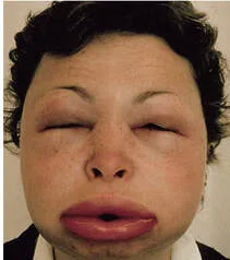
Subcutaneous edema that results in severe deformity if it affects the face.
The complement system is involved in anomalies in the laboratory: low C4 value in the presence of normal C1 and C3 levels, and decreased levels of C1-esterase inhibitor (85%) or malfunctioning inhibitor (15%). Bradykinin development causes angioedema because the main inhibitor of kallikrein and Hageman factor—two enzymes necessary for kinin generation—is C1-esterase inhibitor. Events may pose a hazard to life.
Stanazolol or Danazol. prolonged treatment. Be on the lookout for irregular menses and hirsutism.
Whole fresh plasma or a C1-esterase inhibitor in an acute episode. Icatibant is an extremely potent bradykinin-B2-receptor antagonist that can be used subcutaneously.
a dangerous autosomal dominant condition that could develop after emotional or physical trauma.
Rarely does urticaria occur. The patient presents with acute stomach pain due to angioedema of the intestinal wall, laryngeal edema episodes, and angioedema of the face and limbs.
Subcutaneous edema that results in severe deformity if it affects the face.
The complement system is involved in anomalies in the laboratory: low C4 value in the presence of normal C1 and C3 levels, and decreased levels of C1-esterase inhibitor (85%) or malfunctioning inhibitor (15%). Bradykinin development causes angioedema because the main inhibitor of kallikrein and Hageman factor—two enzymes necessary for kinin generation—is C1-esterase inhibitor. Events may pose a hazard to life.
Stanazolol or Danazol. prolonged treatment. Be on the lookout for irregular menses and hirsutism.
Whole fresh plasma or a C1-esterase inhibitor in an acute episode. Icatibant is an extremely potent bradykinin-B2-receptor antagonist that can be used subcutaneously.
- Published on
Dermatology - Melanoma In Situ (MIS)
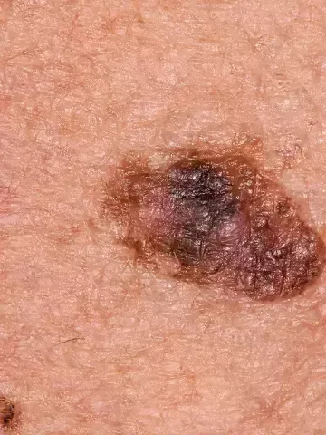
When melanoma cells are restricted to the epidermis, above the basement membrane, MIS is largely employed as a histological definition.
Hyperplasia, atypia, and spread of basilar melanocytic atypia can occur in single file alignment along the basal membrane (lentigo maligna, LM) or dispersed in a pagetoid pattern throughout the epidermis (superficial spreading melanoma, SSM).
The LM or flat SSM clinical correlations of MIS are known. Because LM is flat and lies at the skin's surface, it is either a macule with scarcely noticeable elevation, uneven boundaries, and noticeable color variation, with brown, black, or reddish tones predominating but neither gray nor blue.
The early SSM is a thinly raised plaque with a comparatively uniform border and a remarkable range of colors, including tan, brown, black, and even red or white.
Excision is the method of treatment.
When melanoma cells are restricted to the epidermis, above the basement membrane, MIS is largely employed as a histological definition.
Hyperplasia, atypia, and spread of basilar melanocytic atypia can occur in single file alignment along the basal membrane (lentigo maligna, LM) or dispersed in a pagetoid pattern throughout the epidermis (superficial spreading melanoma, SSM).
The LM or flat SSM clinical correlations of MIS are known. Because LM is flat and lies at the skin's surface, it is either a macule with scarcely noticeable elevation, uneven boundaries, and noticeable color variation, with brown, black, or reddish tones predominating but neither gray nor blue.
The early SSM is a thinly raised plaque with a comparatively uniform border and a remarkable range of colors, including tan, brown, black, and even red or white.
Excision is the method of treatment.
- Published on
Dermatology - Pyoderma Gangrenosum
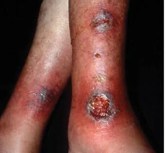
Acute or chronic pyoderma gangrenosum is an idiopathic skin disease that is quite debilitating. Though it can also happen on its own, it frequently coexists with a systemic illness, including arthritis, inflammatory bowel disease, hematologic dyscriasis, and cancer.
The patient can seem unwell. As shown previously, 50% of cases have no accompanying systemic disease and up to 50% have.
A painful superficial hemorrhagic pustule with an erythematous circle surrounds it acutely.
Breakdown happens during the creation of ulcers, which have uneven, elevated, undermined, swampy, and dusky-red or purple borders with pus-draining perforations. The ulcer's base is purulent and has hemorrhagic discharge; necrotic eschar, either with or without granulation tissue, covers portion of the ulcer. A halo of erythema develops centrifugally at the ulcer's spreading edge, and pustules can be detected in the base of the ulcer and at its advancing border. When a lesion is chronic, it may gradually spread over a sizable portion of the body, displaying severe granulation inside the ulcer as well as crusting and even hyperkeratosis around the edges. Lesions are often isolated, however they can also occur in groups and eventually come together. Atrophic cribriform scars are thin and atrophic after ulcer healing. There are hemorrhagic blisters that ulcerate in the bullous form. Massive ulceration of the conjunctiva and oral mucosa occurs infrequently.
Clinical diagnosis and compatible nonspecific dermatopathology demonstrating neutrophilic inflammation with abscesses and necrosis are used to validate the diagnosis. Ecthyma, Ecthyma gangrenosum, clostridial infection, atypical mycobacterial infection, deep mycoses, amebiasis, leishmaniasis, bromoderma, pemphigus vegetans, stasis ulcers, and Wegener granulomatosis are among the conditions included in the differential.
If left untreated, the illness can linger for months or even years, however it can also recover on its own.
Attend to any underlying medical conditions. It could be necessary to use high dosages of oral glucocorticoids or IV glucocorticoid pulse treatment (1-2 g/day prednisolone). Effective treatments include sulfasalazine (especially in Crohn's disease cases), sulfones, cyclosporine, and, more recently, infliximab, etanercept, and adalimumab.
Topical tacrolimus ointment or intralesional triamcinolone may be useful for isolated minor lesions.
Acute or chronic pyoderma gangrenosum is an idiopathic skin disease that is quite debilitating. Though it can also happen on its own, it frequently coexists with a systemic illness, including arthritis, inflammatory bowel disease, hematologic dyscriasis, and cancer.
The patient can seem unwell. As shown previously, 50% of cases have no accompanying systemic disease and up to 50% have.
A painful superficial hemorrhagic pustule with an erythematous circle surrounds it acutely.
Breakdown happens during the creation of ulcers, which have uneven, elevated, undermined, swampy, and dusky-red or purple borders with pus-draining perforations. The ulcer's base is purulent and has hemorrhagic discharge; necrotic eschar, either with or without granulation tissue, covers portion of the ulcer. A halo of erythema develops centrifugally at the ulcer's spreading edge, and pustules can be detected in the base of the ulcer and at its advancing border. When a lesion is chronic, it may gradually spread over a sizable portion of the body, displaying severe granulation inside the ulcer as well as crusting and even hyperkeratosis around the edges. Lesions are often isolated, however they can also occur in groups and eventually come together. Atrophic cribriform scars are thin and atrophic after ulcer healing. There are hemorrhagic blisters that ulcerate in the bullous form. Massive ulceration of the conjunctiva and oral mucosa occurs infrequently.
Clinical diagnosis and compatible nonspecific dermatopathology demonstrating neutrophilic inflammation with abscesses and necrosis are used to validate the diagnosis. Ecthyma, Ecthyma gangrenosum, clostridial infection, atypical mycobacterial infection, deep mycoses, amebiasis, leishmaniasis, bromoderma, pemphigus vegetans, stasis ulcers, and Wegener granulomatosis are among the conditions included in the differential.
If left untreated, the illness can linger for months or even years, however it can also recover on its own.
Attend to any underlying medical conditions. It could be necessary to use high dosages of oral glucocorticoids or IV glucocorticoid pulse treatment (1-2 g/day prednisolone). Effective treatments include sulfasalazine (especially in Crohn's disease cases), sulfones, cyclosporine, and, more recently, infliximab, etanercept, and adalimumab.
Topical tacrolimus ointment or intralesional triamcinolone may be useful for isolated minor lesions.
- Published on
Dermatology - Lymphangioma
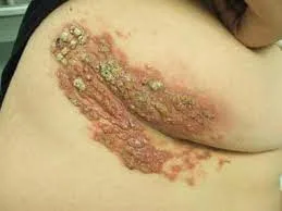
A microcystic lesion known as a lymphphangioma, or lymphatic malformation, can develop throughout infancy or childhood or be present from birth. Rarely, it could vanish on its own. There could be a bacterial infection.
Lymphatic malformations can manifest as solitary lesions that are isolated or as huge patches that span up to 10 by 20 centimeters; they can also be linked to capillary venous lymphatic abnormalities.
Lesions consist of several clusters of tiny, macroscopic vesicles that are filled with clear or serosanguinous fluid (also known as "frog-spawn"). These are microcystic lesions, not genuine vesicles (lymphangioma). On the other hand, macrocystic lesions, or cystic hygromas, manifest as sizable, pliable subcutaneous tumors.
Clinical diagnosis is made.
If possible, the lesion can be removed, or it can be treated with sclerotherapy.
A microcystic lesion known as a lymphphangioma, or lymphatic malformation, can develop throughout infancy or childhood or be present from birth. Rarely, it could vanish on its own. There could be a bacterial infection.
Lymphatic malformations can manifest as solitary lesions that are isolated or as huge patches that span up to 10 by 20 centimeters; they can also be linked to capillary venous lymphatic abnormalities.
Lesions consist of several clusters of tiny, macroscopic vesicles that are filled with clear or serosanguinous fluid (also known as "frog-spawn"). These are microcystic lesions, not genuine vesicles (lymphangioma). On the other hand, macrocystic lesions, or cystic hygromas, manifest as sizable, pliable subcutaneous tumors.
Clinical diagnosis is made.
If possible, the lesion can be removed, or it can be treated with sclerotherapy.
- Published on
Dermatology - Genital Herpes Simplex Virus Infection
A persistent sexually transmitted virus condition called genital herpes is characterized by both symptomatic and silent viral shedding. This is a chronic infection that becomes worse with aging and comes back. Meningitis and radiation injury are prevented by treating the first episode infection. Erythema multiforme, which appears 1-2 weeks following an epidemic, can exacerbate recurrences.
Many people who are infected are unaware that they are infected. When symptomatic, the following symptoms may be present: considerable pain, regional lymphadenopathy, fever, headache, malaise, and myalgia, which peak in the first 2-4 days following the beginning of lesions and resolve in the following 2-4 days. Prior to skin eruption, recurrences may present with itching, burning, fissures, redness, irritation, dysuria, sciatica, and rectal discomfort.
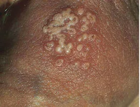
The majority of clinical lesions appear as erosion, "abrasions," or fissures and are actually small breaches in the mucocutaneous epithelium. Erythematous papules that develop into vesicles or pustules are the typical lesion; they eventually erode as the underlying epidermis sloughs off. In 2-4 weeks, lesions resolve; scarring is rare and postinflammatory hypo- or hyperpigmentation is typical. Lesions in patients with impaired immune systems typically have larger areas and take longer to heal. Recurrence lesions are similar, but smaller in size, and they recover in a couple of weeks.
Viral culture is used to confirm the diagnosis, and serology, direct fluorescent antibody (DFA), or both may be necessary. Make sure there is no co-infection with any other STDs.
The differential diagnosis consists of gonococcal erosion, trauma, candidiasis, syphilitic chancre, eruption from a fixed medication, and chancroid.
Encourage patients to abstain from sexual activity if they have lesions and to use condoms while having sex.
Acyclovir 400 mg should be taken five times a day for ten days or until the lesions clear up for the first episode; for recurrences, use 400 mg three times a day for five days or 800 mg three times a day for two days. Famaciclovir 125 mg twice daily for five days or 1 g once daily for five days are the alternative options for valacyclovir 500 mg twice daily for seven days or 1000 mg twice daily for five days. Acyclovir 400 mg twice a day, valcyclovir 500–1000 mg once a day, or famciclovir 250 mg once a day should be administered for suppressive therapy. If you are seriously immunocompromised, have acyclovir or foscarnet intravenously.
A persistent sexually transmitted virus condition called genital herpes is characterized by both symptomatic and silent viral shedding. This is a chronic infection that becomes worse with aging and comes back. Meningitis and radiation injury are prevented by treating the first episode infection. Erythema multiforme, which appears 1-2 weeks following an epidemic, can exacerbate recurrences.
Many people who are infected are unaware that they are infected. When symptomatic, the following symptoms may be present: considerable pain, regional lymphadenopathy, fever, headache, malaise, and myalgia, which peak in the first 2-4 days following the beginning of lesions and resolve in the following 2-4 days. Prior to skin eruption, recurrences may present with itching, burning, fissures, redness, irritation, dysuria, sciatica, and rectal discomfort.
The majority of clinical lesions appear as erosion, "abrasions," or fissures and are actually small breaches in the mucocutaneous epithelium. Erythematous papules that develop into vesicles or pustules are the typical lesion; they eventually erode as the underlying epidermis sloughs off. In 2-4 weeks, lesions resolve; scarring is rare and postinflammatory hypo- or hyperpigmentation is typical. Lesions in patients with impaired immune systems typically have larger areas and take longer to heal. Recurrence lesions are similar, but smaller in size, and they recover in a couple of weeks.
Viral culture is used to confirm the diagnosis, and serology, direct fluorescent antibody (DFA), or both may be necessary. Make sure there is no co-infection with any other STDs.
The differential diagnosis consists of gonococcal erosion, trauma, candidiasis, syphilitic chancre, eruption from a fixed medication, and chancroid.
Encourage patients to abstain from sexual activity if they have lesions and to use condoms while having sex.
Acyclovir 400 mg should be taken five times a day for ten days or until the lesions clear up for the first episode; for recurrences, use 400 mg three times a day for five days or 800 mg three times a day for two days. Famaciclovir 125 mg twice daily for five days or 1 g once daily for five days are the alternative options for valacyclovir 500 mg twice daily for seven days or 1000 mg twice daily for five days. Acyclovir 400 mg twice a day, valcyclovir 500–1000 mg once a day, or famciclovir 250 mg once a day should be administered for suppressive therapy. If you are seriously immunocompromised, have acyclovir or foscarnet intravenously.
- Published on
Dermatology - Angioedema and Urticaria
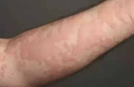
Both urticaria and angioedema are edematous conditions that affect the cutaneous vasculature at the papillary and deep levels. Papillary wheals make up urticaria, while greater edema involving the dermis and subcutaneous tissue is known as angioedema. These could be recurring, chronic, or acute.
The patient may experience paresthesia from carpal tunnel syndrome and neuropathy, as well as signs of hepatic, renal, and GI involvement, fatigue, weakness, anorexia, weight loss, malaise, or dyspnea. Speech, eating, and breathing can all be affected by oral and throat edema. Cold, sun, allergy, immunologic, drug-induced, autoimmune, physically mediated (dermatographism, pressure or vibration generated), and genetic kinds are among the variations.
Damage
Lesions in solar, pressure, vibration, cold urticaria, and angioedema are localized and restricted to the trigger mechanism location. They are typically regional or widespread in nature. Due to confluence and resolution in one place and progression in another, urticaria are finely defined wheals that range in size from less than 1 cm to more than 8 cm, and they can be round, oval, arciform, annular, or serpiginous. The erythematous or white ring is present. Lesions are itchy and fleeting. Angioedema is a skin-colored, momentary swelling caused by subcutaneous edema that can affect the tongue, lips, eyelids, or other parts of the face or limbs.
Clinical diagnosis is made.
Use hydroxyzine, terfenadine, or loratadine, cetirizine, or flufenadine as antihistamines and H1 blockers. The majority of chronic cases are managed with fexofenadine 180 mg/day or loratadine 10–20 mg/day. Recurrences are typically the result of stopping therapy. When anxiety and depression are linked to severe urticaria, doxepin, a tricyclic antidepressant with strong H1 antihistaminic activity, can be helpful.
Use prednisone for acute urticaria associated with edema. If antihistamines have little impact on chronic idiopathic or autoimmune urticaria, switch to cyclosporine and reduce gradually. If glucocorticoids are contraindicated, or if side effects develop, continue using cyclosporine.
Both urticaria and angioedema are edematous conditions that affect the cutaneous vasculature at the papillary and deep levels. Papillary wheals make up urticaria, while greater edema involving the dermis and subcutaneous tissue is known as angioedema. These could be recurring, chronic, or acute.
The patient may experience paresthesia from carpal tunnel syndrome and neuropathy, as well as signs of hepatic, renal, and GI involvement, fatigue, weakness, anorexia, weight loss, malaise, or dyspnea. Speech, eating, and breathing can all be affected by oral and throat edema. Cold, sun, allergy, immunologic, drug-induced, autoimmune, physically mediated (dermatographism, pressure or vibration generated), and genetic kinds are among the variations.
Damage
Lesions in solar, pressure, vibration, cold urticaria, and angioedema are localized and restricted to the trigger mechanism location. They are typically regional or widespread in nature. Due to confluence and resolution in one place and progression in another, urticaria are finely defined wheals that range in size from less than 1 cm to more than 8 cm, and they can be round, oval, arciform, annular, or serpiginous. The erythematous or white ring is present. Lesions are itchy and fleeting. Angioedema is a skin-colored, momentary swelling caused by subcutaneous edema that can affect the tongue, lips, eyelids, or other parts of the face or limbs.
Clinical diagnosis is made.
Use hydroxyzine, terfenadine, or loratadine, cetirizine, or flufenadine as antihistamines and H1 blockers. The majority of chronic cases are managed with fexofenadine 180 mg/day or loratadine 10–20 mg/day. Recurrences are typically the result of stopping therapy. When anxiety and depression are linked to severe urticaria, doxepin, a tricyclic antidepressant with strong H1 antihistaminic activity, can be helpful.
Use prednisone for acute urticaria associated with edema. If antihistamines have little impact on chronic idiopathic or autoimmune urticaria, switch to cyclosporine and reduce gradually. If glucocorticoids are contraindicated, or if side effects develop, continue using cyclosporine.
- Published on
Dermatology - Cherry Angiomas
Cherry angiomas are extremely prevalent, and you can be sure that practically every old person has at least one of them.
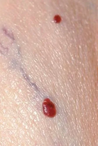
Cherry angiomas are vascular lesions that are dome-shaped and can range from bright red to violaceous or even black in color. As well as being asymptomatic, cherry angiomas can also manifest as numerous tiny red papular patches that mimic petechiae.
It is most common for them to be found on the trunk.
In clinical settings, a diagnosis is made.
Cherry angiomas have little significance other than their appearance from a cosmetic standpoint. For cosmetic purposes, electrocoagulation or laser coagulation may be used as a treatment option. The use of cryosurgery is not successful.
Cherry angiomas are extremely prevalent, and you can be sure that practically every old person has at least one of them.
Cherry angiomas are vascular lesions that are dome-shaped and can range from bright red to violaceous or even black in color. As well as being asymptomatic, cherry angiomas can also manifest as numerous tiny red papular patches that mimic petechiae.
It is most common for them to be found on the trunk.
In clinical settings, a diagnosis is made.
Cherry angiomas have little significance other than their appearance from a cosmetic standpoint. For cosmetic purposes, electrocoagulation or laser coagulation may be used as a treatment option. The use of cryosurgery is not successful.
- Published on
Dermatology - Phytophotodermis
Phytophotodermatitis is an inflammation of the skin that can be brought on by coming into touch with specific plants while being exposed to sunshine for either recreational or occupational purposes (plant + light = dermatitis). In a number of plant groups, the inflammatory response is a phototoxic reaction that occurs in response to compounds that are photosensitizing. Lime, celery, and meadow grass are common causes of the types that are most commonly developed.
A history of exposure to specific plants, such as lime, lemon, wild parsley, celery, giant hogweed, parsnips, carrot greens, and figs, as well as the usage of perfumes that contain oil of bergamot, which contains bergapten, 5-methoxypsoralen, is provided by the patient. Those patients who have been exposed to the perfume or the plant may only acquire streaks of pigmentation in the places where the perfume was applied. The lesions cause a sensation similar to that of a sunburn, alongside pain and, later on, pruritus.
lesions (plural)
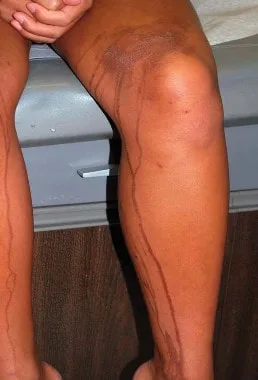
Erythema, edema, vesicles, and bullae are all symptoms that appear suddenly. It's possible that the lesions will look pseudopapular before the vesicles become visible. On the locations of touch, particularly the arms, legs, and face, there are frequently strange streaks and patterns that have been artificially created. Berloque dermatitis, also known as residual hyperpigmentation, is a possibility.
Through the identification of patterns and the meticulous examination of history, a diagnosis can be determined. The differential diagnosis consists mostly of acute irritating contact dermatitis, along with poison ivy dermatitis with a streaky pattern; nonetheless, this is an eczematous condition.
During the acute vesicular stage, it is possible that wet dressings will be required. Topical glucocorticoids are a therapy option for symptomatic conditions. In the event that the offending substance cannot be avoided, the patient should be encouraged to either not expose themselves to the sun or to wear protective clothes.
Phytophotodermatitis is an inflammation of the skin that can be brought on by coming into touch with specific plants while being exposed to sunshine for either recreational or occupational purposes (plant + light = dermatitis). In a number of plant groups, the inflammatory response is a phototoxic reaction that occurs in response to compounds that are photosensitizing. Lime, celery, and meadow grass are common causes of the types that are most commonly developed.
A history of exposure to specific plants, such as lime, lemon, wild parsley, celery, giant hogweed, parsnips, carrot greens, and figs, as well as the usage of perfumes that contain oil of bergamot, which contains bergapten, 5-methoxypsoralen, is provided by the patient. Those patients who have been exposed to the perfume or the plant may only acquire streaks of pigmentation in the places where the perfume was applied. The lesions cause a sensation similar to that of a sunburn, alongside pain and, later on, pruritus.
lesions (plural)
Erythema, edema, vesicles, and bullae are all symptoms that appear suddenly. It's possible that the lesions will look pseudopapular before the vesicles become visible. On the locations of touch, particularly the arms, legs, and face, there are frequently strange streaks and patterns that have been artificially created. Berloque dermatitis, also known as residual hyperpigmentation, is a possibility.
Through the identification of patterns and the meticulous examination of history, a diagnosis can be determined. The differential diagnosis consists mostly of acute irritating contact dermatitis, along with poison ivy dermatitis with a streaky pattern; nonetheless, this is an eczematous condition.
During the acute vesicular stage, it is possible that wet dressings will be required. Topical glucocorticoids are a therapy option for symptomatic conditions. In the event that the offending substance cannot be avoided, the patient should be encouraged to either not expose themselves to the sun or to wear protective clothes.