- Published on
Dermatology - Nail Psoriasis
The most prevalent dermatosis that affects the nail apparatus is psoriasis, which affects the nails in >50% of cases, up to 80–90% of cases over the course of a lifetime.
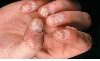
Elkonyxis, also known as pitting, will be present in the nail matrix. These small, shallow punctate depressions come in a variety of sizes and shapes. Though they can also occur as regular lines (transverse; long axis) or in grid-like patterns, their characteristics are isolation and depth.
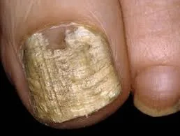
Lesions in the toenails are rare. The nail is rough, brittle, and dull. Proximal nail matrix destruction is linked to sandpaper nails, or twenty-nail dystrophy; however, this is general and can also be observed in alopecia areata, lichen planus, and atopic dermatitis.
Repeated transverse depressions may resemble the "washboard" nails of tic habits, where the cuticle is pushed back.
Longitudinal ridging, akin to melted wax, could exist. One or two millimeter white spots in the nail plate called punctate leukonychia are sometimes misdiagnosed as injuries.
An oval, salmon-colored nail bed called "oil" patches may be present, and onycholysis may develop as a result, affecting the hyponychium medially or laterally.
There could be secondary infections. Subungual hyperkeratosis, or raising of the nail plate off the hyponychium, is possible.
A clinical exam and history are used to make a diagnosis. Onycholysis, onychomycosis, trauma (toenail), eczema, and alopecia areata are among the differential diagnoses.
Intralesional triamcinolone 3-5 mg/mL may be useful for matrix involvement. Topical steroid (occluded) decreases hyperkeratosis in nail bed psoriasis. Nail apparatus psoriasis is frequently improved by systemic therapy, such as methotrexate, acitretin, or "biologics," though results may not show up for some months after treatment has finished.
The most prevalent dermatosis that affects the nail apparatus is psoriasis, which affects the nails in >50% of cases, up to 80–90% of cases over the course of a lifetime.
Elkonyxis, also known as pitting, will be present in the nail matrix. These small, shallow punctate depressions come in a variety of sizes and shapes. Though they can also occur as regular lines (transverse; long axis) or in grid-like patterns, their characteristics are isolation and depth.
Lesions in the toenails are rare. The nail is rough, brittle, and dull. Proximal nail matrix destruction is linked to sandpaper nails, or twenty-nail dystrophy; however, this is general and can also be observed in alopecia areata, lichen planus, and atopic dermatitis.
Repeated transverse depressions may resemble the "washboard" nails of tic habits, where the cuticle is pushed back.
Longitudinal ridging, akin to melted wax, could exist. One or two millimeter white spots in the nail plate called punctate leukonychia are sometimes misdiagnosed as injuries.
An oval, salmon-colored nail bed called "oil" patches may be present, and onycholysis may develop as a result, affecting the hyponychium medially or laterally.
There could be secondary infections. Subungual hyperkeratosis, or raising of the nail plate off the hyponychium, is possible.
A clinical exam and history are used to make a diagnosis. Onycholysis, onychomycosis, trauma (toenail), eczema, and alopecia areata are among the differential diagnoses.
Intralesional triamcinolone 3-5 mg/mL may be useful for matrix involvement. Topical steroid (occluded) decreases hyperkeratosis in nail bed psoriasis. Nail apparatus psoriasis is frequently improved by systemic therapy, such as methotrexate, acitretin, or "biologics," though results may not show up for some months after treatment has finished.
- Published on
Dermatology - Drug Induced Pustular Rash
Pustular Rash Caused by Drug (Adverse Cutaneous Drug Reactions)
Acute generalized exanthematous pustulosis (AGEP) is a febrile eruption with an estimated yearly frequency of 1-2 million cases. It is frequently linked to leukocytosis. Skin lesions may not show for one to three weeks following drug administration; in patients who have already been sensitized, skin signs may manifest in two to three days.
Elevated blood neutrophils and fever are frequent. After less than 15 days, there is spontaneous resolve, which is followed by generalized desquamation around two weeks later.
Damage
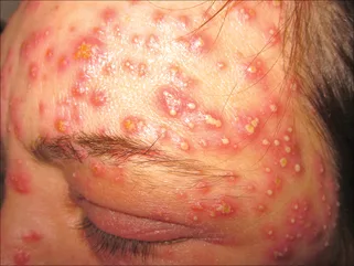
Nonfollicular sterile pustules can be randomly distributed or aggregated, typically beginning in the skin folds and/or on the face, and they appear on a diffuse, edematous erythema.
Clinical diagnosis and differential diagnosis include subcorneal pustular dermatosis (Sneddon-Wilkinson disease), pustular psoriasis, and the hypersensitivity syndrome reaction with pustulation. Iodides, bromides, adrenocorticotropic hormone (ACTH), glucocorticoids, isoniazid, androgens, lithium, actinomycin D, and phenytoin are linked to acneiform pustular eruptions. The EGFR tyrosine kinase inhibitors erlotinib, gefitinib, cetuximab, and panitumumab cause non-acneiform pustules that typically appear on the face but can also occur in unexpected locations including the arms and legs. These pustules are also typically monomorphous. Usually, comedones are not present.
Stop using the offending medication and give the patient supportive treatment.
Pustular Rash Caused by Drug (Adverse Cutaneous Drug Reactions)
Acute generalized exanthematous pustulosis (AGEP) is a febrile eruption with an estimated yearly frequency of 1-2 million cases. It is frequently linked to leukocytosis. Skin lesions may not show for one to three weeks following drug administration; in patients who have already been sensitized, skin signs may manifest in two to three days.
Elevated blood neutrophils and fever are frequent. After less than 15 days, there is spontaneous resolve, which is followed by generalized desquamation around two weeks later.
Damage
Nonfollicular sterile pustules can be randomly distributed or aggregated, typically beginning in the skin folds and/or on the face, and they appear on a diffuse, edematous erythema.
Clinical diagnosis and differential diagnosis include subcorneal pustular dermatosis (Sneddon-Wilkinson disease), pustular psoriasis, and the hypersensitivity syndrome reaction with pustulation. Iodides, bromides, adrenocorticotropic hormone (ACTH), glucocorticoids, isoniazid, androgens, lithium, actinomycin D, and phenytoin are linked to acneiform pustular eruptions. The EGFR tyrosine kinase inhibitors erlotinib, gefitinib, cetuximab, and panitumumab cause non-acneiform pustules that typically appear on the face but can also occur in unexpected locations including the arms and legs. These pustules are also typically monomorphous. Usually, comedones are not present.
Stop using the offending medication and give the patient supportive treatment.
- Published on
Dermatology - Candidal Infection of the Nail
Nail infection caused by Candida
Candida spp. infections of the nail can cause the immune-compromised host's nail to fall off. Rarely are environmental fungus primary infections; instead, they produce secondary colonization of damaged nails.
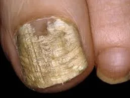
Nail candidiasis can result in a persistent, severe, and sporadic infection of the paronychia, accompanied by erythema, pus, and discomfort. The nail may develop transverse furrows, areas of opacification, and discolorations of white, yellow, green, or black as it becomes dystrophic. Diabetes patients may develop subungual candidiasis and an onycholytic space abscess. Children with HIV/AIDS frequently develop paronychia and onychia, which are frequently linked to mucosal candidiasis.
Tinea unguium, psoriasis, eczema, chronic paronychia, and lichen planus are among the differential diagnoses made using direct microscopy and potassium hydroxide.
Use oral antifungal medications to treat.
Nail infection caused by Candida
Candida spp. infections of the nail can cause the immune-compromised host's nail to fall off. Rarely are environmental fungus primary infections; instead, they produce secondary colonization of damaged nails.
Nail candidiasis can result in a persistent, severe, and sporadic infection of the paronychia, accompanied by erythema, pus, and discomfort. The nail may develop transverse furrows, areas of opacification, and discolorations of white, yellow, green, or black as it becomes dystrophic. Diabetes patients may develop subungual candidiasis and an onycholytic space abscess. Children with HIV/AIDS frequently develop paronychia and onychia, which are frequently linked to mucosal candidiasis.
Tinea unguium, psoriasis, eczema, chronic paronychia, and lichen planus are among the differential diagnoses made using direct microscopy and potassium hydroxide.
Use oral antifungal medications to treat.
- Published on
Dermatology - Tinea Pedis
Tinea Pedis
A particular class of fungus known as dermatiophytes is responsible for the infection of keratinized cutaneous tissues that causes tinea; arthrospores from these species can live for up to a year in skin scales.
The most prevalent ways for transmission are from person to person, from animals, and, less frequently, via soil. The majority of tinea pedis cases start in infancy or early adulthood. The environment, occlusive shoes, and hyperhidrosis are risk factors.
The duration of tinea pedis can range from months to years or even a lifetime. It is frequently accompanied by a history of tinea unguium in the toenails and is associated with tinea in other locations. Usually asymptomatic, pruritus can occur. Bacterial infections can recur later.
Damage
The lesions are macerated, well-defined, erythematous, scaling, and may or may not have bulla.
There may be bulla development or fissuring in the interdigital space. There are tiny papules on the edge, fine white scaling, and hyperkeratosis on the remaining portion of the foot. Any pattern can affect one or both feet, but bilateral involvement is more typical.
In the event of a subsequent infection or if interdigital tinea spreads to the plantar and lateral foot, ulceration may result.
Fungal hyphae can be seen by direct microscopy of skin scrapings that have been taken using a no. 15 scalpel blade, the edge of a glass microscope slide, or a toothbrush (cervical or tooth brush) and covered with a drop of potassium hydroxide (KOH). Erythasma, pitted keratolysis, psoriasis, and eczematous dermatitis (dyshidrotic, atopic, allergic contact) are among the conditions included in the differential.
Use imidazole powder, benzoyl peroxide wash, and dry skin to stop recurrences. Take care of any concurrent nail infections.
Use allylamine lotion (naftifin, terbinafine); naphthionate ointment (tolnaftate); imidazole creams (clotrimazole, miconazole, ketoconazole, econazole, oxiconizole, suconizole, sertaconazole); or substituted pyridine (ciclopirox olamine).
Among oral antidermophytic agents, Systemic Terbinafine 250-mg tablet is the most effective allylamine. Fluconazole 100-, 150-, or 200-mg pills or oral suspension (10 or 40 mg/mL) are substitutes for itraconazole 100-mg capsules or oral solution (10 mg/mL).
Tinea Pedis
A particular class of fungus known as dermatiophytes is responsible for the infection of keratinized cutaneous tissues that causes tinea; arthrospores from these species can live for up to a year in skin scales.
The most prevalent ways for transmission are from person to person, from animals, and, less frequently, via soil. The majority of tinea pedis cases start in infancy or early adulthood. The environment, occlusive shoes, and hyperhidrosis are risk factors.
The duration of tinea pedis can range from months to years or even a lifetime. It is frequently accompanied by a history of tinea unguium in the toenails and is associated with tinea in other locations. Usually asymptomatic, pruritus can occur. Bacterial infections can recur later.
Damage
The lesions are macerated, well-defined, erythematous, scaling, and may or may not have bulla.
There may be bulla development or fissuring in the interdigital space. There are tiny papules on the edge, fine white scaling, and hyperkeratosis on the remaining portion of the foot. Any pattern can affect one or both feet, but bilateral involvement is more typical.
In the event of a subsequent infection or if interdigital tinea spreads to the plantar and lateral foot, ulceration may result.
Fungal hyphae can be seen by direct microscopy of skin scrapings that have been taken using a no. 15 scalpel blade, the edge of a glass microscope slide, or a toothbrush (cervical or tooth brush) and covered with a drop of potassium hydroxide (KOH). Erythasma, pitted keratolysis, psoriasis, and eczematous dermatitis (dyshidrotic, atopic, allergic contact) are among the conditions included in the differential.
Use imidazole powder, benzoyl peroxide wash, and dry skin to stop recurrences. Take care of any concurrent nail infections.
Use allylamine lotion (naftifin, terbinafine); naphthionate ointment (tolnaftate); imidazole creams (clotrimazole, miconazole, ketoconazole, econazole, oxiconizole, suconizole, sertaconazole); or substituted pyridine (ciclopirox olamine).
Among oral antidermophytic agents, Systemic Terbinafine 250-mg tablet is the most effective allylamine. Fluconazole 100-, 150-, or 200-mg pills or oral suspension (10 or 40 mg/mL) are substitutes for itraconazole 100-mg capsules or oral solution (10 mg/mL).
- Published on
Dermatology - Scurvy
Scurvy is an acute or chronic illness that arises in newborns or children who exclusively consume processed milk or in adults who avoid salads and raw vegetables because of a dietary shortage of ascorbic acid (vitamin C). Pregnancy, lactation, and thyrotoxicosis—which is most prevalent in alcoholism—are precipitating factors.
After one to three months without vitamin C, lassitude, weakness, arthralgia, and myalgia are the symptoms of scurvy. A bleeding injury into the joints and periosteum of long bones causes excruciating swellings and, in children, epiphyseal separation. The rib borders are elevated and the sternum inclines inward.
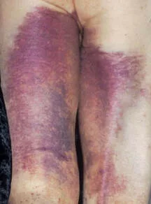
Particularly on the lower legs, petechiae and follicular hyperkeratosis with perifollicular bleeding are lesions that can arise. These perifollicular hyperkeratotic papules, also known as corkscrew hairs, break up and bury the hair, and there are widespread, generally spread ecchymoses.
Splinter hemorrhages occur in nails. The gingiva are spongy, purple, enlarged, and prone to bleeding. Loss of teeth and tooth loosening are possible.
When serum ascorbic acid levels are nil, the diagnosis of macrocytic, normochromic, and normocytic anemia is made. Findings from X-rays are also diagnostic.
If left untreated, scurvy is lethal. When 4 g of ascorbic acid is administered, 100 mg/d is curative in a matter of days to weeks.
Scurvy is an acute or chronic illness that arises in newborns or children who exclusively consume processed milk or in adults who avoid salads and raw vegetables because of a dietary shortage of ascorbic acid (vitamin C). Pregnancy, lactation, and thyrotoxicosis—which is most prevalent in alcoholism—are precipitating factors.
After one to three months without vitamin C, lassitude, weakness, arthralgia, and myalgia are the symptoms of scurvy. A bleeding injury into the joints and periosteum of long bones causes excruciating swellings and, in children, epiphyseal separation. The rib borders are elevated and the sternum inclines inward.
Particularly on the lower legs, petechiae and follicular hyperkeratosis with perifollicular bleeding are lesions that can arise. These perifollicular hyperkeratotic papules, also known as corkscrew hairs, break up and bury the hair, and there are widespread, generally spread ecchymoses.
Splinter hemorrhages occur in nails. The gingiva are spongy, purple, enlarged, and prone to bleeding. Loss of teeth and tooth loosening are possible.
When serum ascorbic acid levels are nil, the diagnosis of macrocytic, normochromic, and normocytic anemia is made. Findings from X-rays are also diagnostic.
If left untreated, scurvy is lethal. When 4 g of ascorbic acid is administered, 100 mg/d is curative in a matter of days to weeks.
- Published on
Dermatology - Mammary Paget Disease
Mammary Paget disease is a malignant tumor that mimics a persistent eczematous dermatitis and affects the nipple or areola unilaterally. It typically affects women over 50 and is indicative of the continuous spread of underlying intraductal carcinoma of the breast, which accounts for 1-4 percent of breast cancer cases. Men are not without uncommon examples. It develops slowly over a few months or years.
Itching, soreness, burning, discharge, bleeding, ulceration, and nipple invasion are possible symptoms.
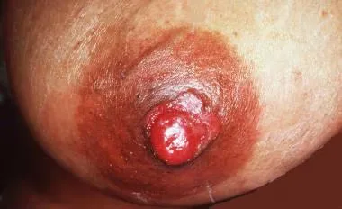
Skin lesions are oval, red, scaling plaques that may be bilateral in nature and have sharp margins. The surface is wet and dripping once the scale is removed.
The size of lesions varies from 0.3 to 15 cm. Early on, the plaque is not indurated; subsequently, it becomes indurated and infiltrated, and nodules may be felt in the breast. Less than half of patients have a palpable underlying breast lump at initial presentation.
When there is a palpable mass beneath the lesion, lymph node metastases happen more frequently.
A biopsy that reveals cancerous cells in the epidermis that are spreading in a pathognomonic way confirms the diagnosis. Use mammography to identify underlying intraductal cancer.
The differential diagnosis consists of squamous cell carcinoma in situ, hereditary pemphigus, nippleareola retention hyperkeratosis, psoriasis, eczematous dermatitis, and benign ductal papilloma. The majority of the time, bilateral nipple eczematous dermatitis is non-induring and reacts quickly to topical glucocorticoids. However, if the "eczema" lasts longer than three weeks, be on the lookout for Paget illness.
Treatment options include surgery, radiation, and/or chemotherapy, much like with other types of breast cancer. If palpable regional nodes are found, dissect the lymph nodes. The prognosis differs. 92% of patients survive 5 years following excision if the breast lump is not perceptible, and 82% survive 10 years.
Of those with palpable breast mass, 38% live for five years and 22% for ten. When lymphadenopathy is present, the prognosis is worse.
Mammary Paget disease is a malignant tumor that mimics a persistent eczematous dermatitis and affects the nipple or areola unilaterally. It typically affects women over 50 and is indicative of the continuous spread of underlying intraductal carcinoma of the breast, which accounts for 1-4 percent of breast cancer cases. Men are not without uncommon examples. It develops slowly over a few months or years.
Itching, soreness, burning, discharge, bleeding, ulceration, and nipple invasion are possible symptoms.
Skin lesions are oval, red, scaling plaques that may be bilateral in nature and have sharp margins. The surface is wet and dripping once the scale is removed.
The size of lesions varies from 0.3 to 15 cm. Early on, the plaque is not indurated; subsequently, it becomes indurated and infiltrated, and nodules may be felt in the breast. Less than half of patients have a palpable underlying breast lump at initial presentation.
When there is a palpable mass beneath the lesion, lymph node metastases happen more frequently.
A biopsy that reveals cancerous cells in the epidermis that are spreading in a pathognomonic way confirms the diagnosis. Use mammography to identify underlying intraductal cancer.
The differential diagnosis consists of squamous cell carcinoma in situ, hereditary pemphigus, nippleareola retention hyperkeratosis, psoriasis, eczematous dermatitis, and benign ductal papilloma. The majority of the time, bilateral nipple eczematous dermatitis is non-induring and reacts quickly to topical glucocorticoids. However, if the "eczema" lasts longer than three weeks, be on the lookout for Paget illness.
Treatment options include surgery, radiation, and/or chemotherapy, much like with other types of breast cancer. If palpable regional nodes are found, dissect the lymph nodes. The prognosis differs. 92% of patients survive 5 years following excision if the breast lump is not perceptible, and 82% survive 10 years.
Of those with palpable breast mass, 38% live for five years and 22% for ten. When lymphadenopathy is present, the prognosis is worse.
- Published on
Dermatology - Cryoglobulinemia
Serum immunoglobulin complexed with other immunoglobulins or proteins (which precipitates at low temperatures and redissolves at 37°C) is known as cryoglobulinemia. Cryoglobinemia is linked to a number of diseases, such as connective tissue diseases, autoimmune diseases, multiple myeloma, Waldenström macroglobulinemia, B cell lymphoma, chronic lymphocytic leukemia, rheumatoid arthritis, systemic lupus erythematosus, Sjögren syndrome, and viral or bacterial infections.
Less than 50% of cases have cold sensitivity. After being exposed to the cold, one may have chills, fever, dyspnea, or diarrhea. Purpura may also occur after extended standing or sitting. Arthralgia, renal symptoms, neurologic symptoms, stomach discomfort, or arterial thrombosis may be caused by involvement of other organ systems. 30% to 60% of people with essential mixed cryoglobinemia go on to experience hypertension, edema, or renal failure as a result of renal illness. Peripheral sensory polyneuropathy, which presents as paresthesias or foot drop, is a sign of neurologic involvement. Hepatosplenomegaly and arthritis could be present.
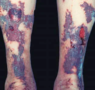
Noninflammatory purpura develops at cold-exposed areas, like the nose tip or the ear helix. It is possible to witness acrocyanosis and Raynaud phenomenon, either with or without serious gangrene resulting in the fingertips, toes, or other areas of the arms and legs. Due to hypersensitivity vasculitis, there may be palpable purpura with bullae and necroses; these are brought on by standing up or being chilly, and they appear as crops on the lower extremities that extend to the thighs and abdomen. Most cases of livedo reticularis occur in the upper and lower extremities. Purpura may be accompanied by cold-induced urticaria.
Cryoglobulin levels and the underlying illness diagnosis validate the diagnosis.
Address the underlying illness.
Serum immunoglobulin complexed with other immunoglobulins or proteins (which precipitates at low temperatures and redissolves at 37°C) is known as cryoglobulinemia. Cryoglobinemia is linked to a number of diseases, such as connective tissue diseases, autoimmune diseases, multiple myeloma, Waldenström macroglobulinemia, B cell lymphoma, chronic lymphocytic leukemia, rheumatoid arthritis, systemic lupus erythematosus, Sjögren syndrome, and viral or bacterial infections.
Less than 50% of cases have cold sensitivity. After being exposed to the cold, one may have chills, fever, dyspnea, or diarrhea. Purpura may also occur after extended standing or sitting. Arthralgia, renal symptoms, neurologic symptoms, stomach discomfort, or arterial thrombosis may be caused by involvement of other organ systems. 30% to 60% of people with essential mixed cryoglobinemia go on to experience hypertension, edema, or renal failure as a result of renal illness. Peripheral sensory polyneuropathy, which presents as paresthesias or foot drop, is a sign of neurologic involvement. Hepatosplenomegaly and arthritis could be present.
Noninflammatory purpura develops at cold-exposed areas, like the nose tip or the ear helix. It is possible to witness acrocyanosis and Raynaud phenomenon, either with or without serious gangrene resulting in the fingertips, toes, or other areas of the arms and legs. Due to hypersensitivity vasculitis, there may be palpable purpura with bullae and necroses; these are brought on by standing up or being chilly, and they appear as crops on the lower extremities that extend to the thighs and abdomen. Most cases of livedo reticularis occur in the upper and lower extremities. Purpura may be accompanied by cold-induced urticaria.
Cryoglobulin levels and the underlying illness diagnosis validate the diagnosis.
Address the underlying illness.
- Published on
Dermatology - Molluscum Contagiosum
A self-limiting viral infection of the epidermis, molluscum contagiosum heals on its own—with the exception of immunocompromised people. Males are more likely than females to contract molluscum infections, and they are more common in children and sexually active adults.
Any location, particularly those that are naturally blocked like the axillae, antecubital, popliteal fossae, and anogenital folds, might become infected. Lesions are propagated by autoinoculation. Mollusca may be found in large quantities in atopic dermatitis areas. Adults with STIs may experience symptoms in the lower belly, thighs, groins, and genitalia. One-sided conjunctivitis can result from mollusca in the conjunctiva.
Damage
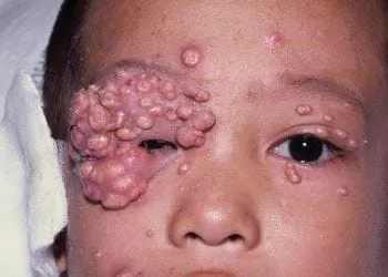
Confluent mosaic plaques can form from skin-colored, round, oval, hemispherical, single or multiple, distributed discrete papules, nodules, or tumors with a central umbilication or depression. Larger lesions may have a central keratotic block, which can be gently extruded to give the lesion a center dimple or umbilication. An inflammatory halo surrounds the mollusk, signaling the start of spontaneous regression, as a result of the host immune system's reaction to the viral antigen. Significant postinflammatory hyperpigmentation in individuals with dark skin tones may arise following therapy or as a result of spontaneous regression.
Clinical diagnosis is made. Human papillomavirus (HPV), condylomata acuminatum, syringoma, keratoacanthoma, squamous cell carcinoma, basal cell carcinoma, and epidermal inclusion cysts are among the conditions included in the differential.
Mollusca self-resolve, hence no treatment is necessary. Office-based treatments include curettage, cryosurgery, and electrodessication if desired for cosmetic impact. Imiquimod 5% cream might work as well.
A self-limiting viral infection of the epidermis, molluscum contagiosum heals on its own—with the exception of immunocompromised people. Males are more likely than females to contract molluscum infections, and they are more common in children and sexually active adults.
Any location, particularly those that are naturally blocked like the axillae, antecubital, popliteal fossae, and anogenital folds, might become infected. Lesions are propagated by autoinoculation. Mollusca may be found in large quantities in atopic dermatitis areas. Adults with STIs may experience symptoms in the lower belly, thighs, groins, and genitalia. One-sided conjunctivitis can result from mollusca in the conjunctiva.
Damage
Confluent mosaic plaques can form from skin-colored, round, oval, hemispherical, single or multiple, distributed discrete papules, nodules, or tumors with a central umbilication or depression. Larger lesions may have a central keratotic block, which can be gently extruded to give the lesion a center dimple or umbilication. An inflammatory halo surrounds the mollusk, signaling the start of spontaneous regression, as a result of the host immune system's reaction to the viral antigen. Significant postinflammatory hyperpigmentation in individuals with dark skin tones may arise following therapy or as a result of spontaneous regression.
Clinical diagnosis is made. Human papillomavirus (HPV), condylomata acuminatum, syringoma, keratoacanthoma, squamous cell carcinoma, basal cell carcinoma, and epidermal inclusion cysts are among the conditions included in the differential.
Mollusca self-resolve, hence no treatment is necessary. Office-based treatments include curettage, cryosurgery, and electrodessication if desired for cosmetic impact. Imiquimod 5% cream might work as well.
- Published on
Dermatology - Trichotillomania
Patients with trichotillomania may deny pulling or plucking their own hair. Trichotillomania is a compulsive desire or habit to pluck hair that may be related to a specific event or persistent stress.
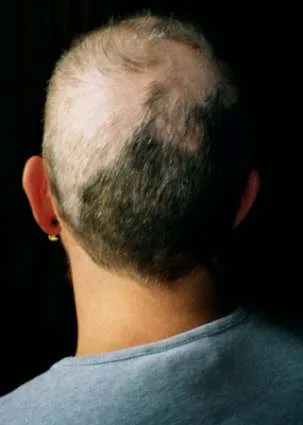
Hair loss can occur on the scalp or in any area that has hair, such as the beard. There is a convergence of small patches of baldness, normal scalp area, and extremely short sparse hairs. Hair loss is typically more noticeable on the dominant hand's side and can coexist with neurotic episodes brought on by forceful tweezer plucking. There are anagen hairs or bluntly broken hairs under the microscope.Clinical diagnosis is determined only after all other potential causes of hair loss have been ruled out.
Careful differential diagnosis is the first step in managing obsessive-compulsive disorder, depression, or other underlying psychiatric issues. This is followed by psychiatric diagnosis and treatment.
Patients with trichotillomania may deny pulling or plucking their own hair. Trichotillomania is a compulsive desire or habit to pluck hair that may be related to a specific event or persistent stress.
Hair loss can occur on the scalp or in any area that has hair, such as the beard. There is a convergence of small patches of baldness, normal scalp area, and extremely short sparse hairs. Hair loss is typically more noticeable on the dominant hand's side and can coexist with neurotic episodes brought on by forceful tweezer plucking. There are anagen hairs or bluntly broken hairs under the microscope.Clinical diagnosis is determined only after all other potential causes of hair loss have been ruled out.
Careful differential diagnosis is the first step in managing obsessive-compulsive disorder, depression, or other underlying psychiatric issues. This is followed by psychiatric diagnosis and treatment.
- Published on
Dermatology - Acanthosis Nigricans
Acanthosis nigricans is a cutaneous sign associated with medication administration, cancer, and endocrine problems (especially diabetes); it has a slow beginning, which accelerates with malignancy. Type 1: An endocrine illness is not related with a hereditary benign. Type 2: Benign has hyperandrogenic conditions, acromegaly/gigantism, Cushing disease, hypogonadal syndromes, Addison disease, hypothyroidism, and insulin-resistant type II diabetes mellitus. Type 3: Pseudo is more common in patients with darker pigmentation and metabolic syndrome, and it is linked to obesity. The result of obesity is insulin resistance. Type 4: Drug-induced conditions can be brought on by excessive dosages of nicotinic acid, growth hormone therapy, glucocorticoid therapy, diethylstilbestrol/oral contraceptives, andstilbestrol in young males. Type 5: Malignant is brought on by a neoplastic illness, most frequently gastrointestinal or genitourinary tract adenocarcinoma; lymphoma or bronchial carcinoma are less frequent causes.
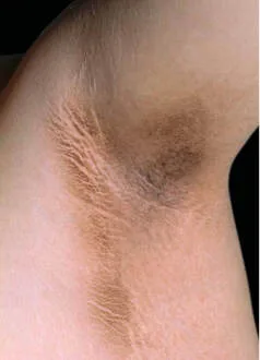
Pigmentation darkening; skin gets thicker, looks unclean, and has more defined wrinkles. The surface gets mangled and rugose. In Type 3, skin tags in body folds and the neck are frequently present, along with a velvety patch on the inner, upper thigh where chafing occurs. Type 5 affects the oral mucosa, the vermilion border of the lips, and exhibits hyperkeratosis and hyperpigmentation. There is hyperkeratosis of the palms and soles, accentuation of the papillary markings (also known as "tripe hands"), involvement of the oral mucosa, and a red border around the lips. Lesions most frequently occur on the knuckles, submammary area, umbilicus, axillae, neck, groin, anogenitalia, and antecubital fossae. The mammillar regions, palms, perioral, and periocular areas are similarly impacted by type 5. The mucocutaneous connections are often affected by type 5, and the oral mucosa has a velvety texture with fine furrows.
The differential includes nicotinic acid intake, X-linked ichthyosis, reticulated papillomatosis (Gougerot-Carteaud syndrome), pityriasis versicolor, and retention hyperkeratosis. The diagnosis is clinical.
Address the related disease. Although they are not particularly effective, topical keratolytic agents and/or topical or systemic retinoids may help skin lesions.
Acanthosis nigricans is a cutaneous sign associated with medication administration, cancer, and endocrine problems (especially diabetes); it has a slow beginning, which accelerates with malignancy. Type 1: An endocrine illness is not related with a hereditary benign. Type 2: Benign has hyperandrogenic conditions, acromegaly/gigantism, Cushing disease, hypogonadal syndromes, Addison disease, hypothyroidism, and insulin-resistant type II diabetes mellitus. Type 3: Pseudo is more common in patients with darker pigmentation and metabolic syndrome, and it is linked to obesity. The result of obesity is insulin resistance. Type 4: Drug-induced conditions can be brought on by excessive dosages of nicotinic acid, growth hormone therapy, glucocorticoid therapy, diethylstilbestrol/oral contraceptives, andstilbestrol in young males. Type 5: Malignant is brought on by a neoplastic illness, most frequently gastrointestinal or genitourinary tract adenocarcinoma; lymphoma or bronchial carcinoma are less frequent causes.
Pigmentation darkening; skin gets thicker, looks unclean, and has more defined wrinkles. The surface gets mangled and rugose. In Type 3, skin tags in body folds and the neck are frequently present, along with a velvety patch on the inner, upper thigh where chafing occurs. Type 5 affects the oral mucosa, the vermilion border of the lips, and exhibits hyperkeratosis and hyperpigmentation. There is hyperkeratosis of the palms and soles, accentuation of the papillary markings (also known as "tripe hands"), involvement of the oral mucosa, and a red border around the lips. Lesions most frequently occur on the knuckles, submammary area, umbilicus, axillae, neck, groin, anogenitalia, and antecubital fossae. The mammillar regions, palms, perioral, and periocular areas are similarly impacted by type 5. The mucocutaneous connections are often affected by type 5, and the oral mucosa has a velvety texture with fine furrows.
The differential includes nicotinic acid intake, X-linked ichthyosis, reticulated papillomatosis (Gougerot-Carteaud syndrome), pityriasis versicolor, and retention hyperkeratosis. The diagnosis is clinical.
Address the related disease. Although they are not particularly effective, topical keratolytic agents and/or topical or systemic retinoids may help skin lesions.