- Published on
Infectious Diseases and Microbiology - Mycetoma ( Madura Foot) ( various saprophytic fungi in soil)
Infectious Diseases and Microbiology - Mycetoma ( Madura Foot) ( various saprophytic fungi in soil)
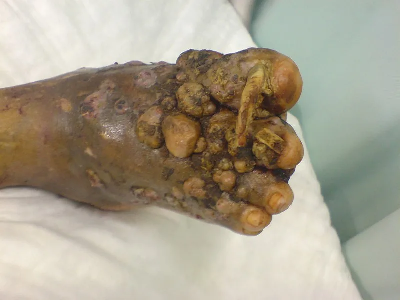
Mycetoma is a medical condition characterized by the formation of a chronic granulomatous infection in the skin and underlying tissues.
Mycetoma, commonly known as Madura foot, is a persistent infection of the subcutaneous tissue caused by various saprophytic fungus found in the soil. Mycetoma agents are widely distributed and spread by inoculation, typically occurring in individuals who go barefoot and sustain injuries. Mycetoma is prevalent in tropical regions but rare in the United States.
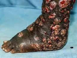
Mycetoma is a condition that is distinguished by the development of abscesses in a specific area, along with swelling, hardening of the tissue, and the presence of sinuses that release fluid. This condition typically occurs in the foot following a traumatic injury. Granulomas have the ability to spread to muscle and bone, causing abnormalities.
The mycetoma granulomatous lesions comprise sclerotia, which are compact masses of hyphae, sometimes known as granules or grains.
The diagnosis is established through the macroscopic detection of granules derived from sclerotia. The dimensions, form, and hue of the granules aid in discerning the causative factor.
The treatment involves surgical debridement, which is often accompanied by the administration of antifungal medications, depending on the specific causative agent. Wearing shoes is a precautionary measure.
Mycetoma is a medical condition characterized by the formation of a chronic granulomatous infection in the skin and underlying tissues.
Mycetoma, commonly known as Madura foot, is a persistent infection of the subcutaneous tissue caused by various saprophytic fungus found in the soil. Mycetoma agents are widely distributed and spread by inoculation, typically occurring in individuals who go barefoot and sustain injuries. Mycetoma is prevalent in tropical regions but rare in the United States.
Mycetoma is a condition that is distinguished by the development of abscesses in a specific area, along with swelling, hardening of the tissue, and the presence of sinuses that release fluid. This condition typically occurs in the foot following a traumatic injury. Granulomas have the ability to spread to muscle and bone, causing abnormalities.
The mycetoma granulomatous lesions comprise sclerotia, which are compact masses of hyphae, sometimes known as granules or grains.
The diagnosis is established through the macroscopic detection of granules derived from sclerotia. The dimensions, form, and hue of the granules aid in discerning the causative factor.
The treatment involves surgical debridement, which is often accompanied by the administration of antifungal medications, depending on the specific causative agent. Wearing shoes is a precautionary measure.
- Published on
Infectious Diseases and Microbiology - Rhinosporidiosis ( Rhinosporidium seeberi)
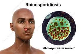
Rhinosporidium seeberi is the causative agent of rhinosporidiosis, which is linked to traumatizing vaccinations and diving. Fish are rhinosporidiosis's natural reservoir. The majority of instances are recorded from Sri Lanka and India.
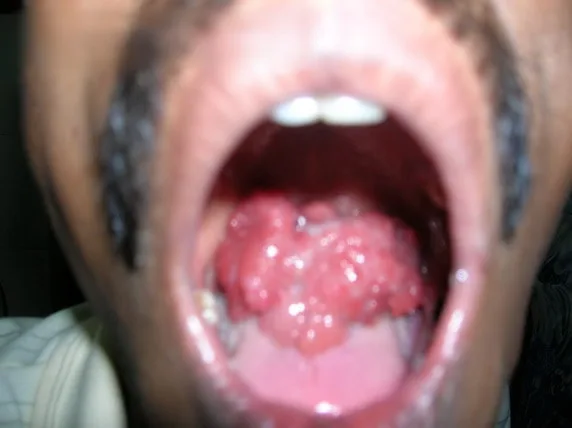
The formation of painless, pedunculated nasal polyps is a characteristic of rhinosporidiosis.
The hallmarks of rhinosporidiosis lesions include an inflammatory cell infiltration, tissue necrosis, and the formation of abscesses.
Large spherules and endospores are seen upon direct microscopic examination of removed tissue or nasal discharge in a KOH preparation.
Amphotericin B injections locally or surgical excision are the two methods used to treat rhinosporidiosis.
Rhinosporidium seeberi is the causative agent of rhinosporidiosis, which is linked to traumatizing vaccinations and diving. Fish are rhinosporidiosis's natural reservoir. The majority of instances are recorded from Sri Lanka and India.
The formation of painless, pedunculated nasal polyps is a characteristic of rhinosporidiosis.
The hallmarks of rhinosporidiosis lesions include an inflammatory cell infiltration, tissue necrosis, and the formation of abscesses.
Large spherules and endospores are seen upon direct microscopic examination of removed tissue or nasal discharge in a KOH preparation.
Amphotericin B injections locally or surgical excision are the two methods used to treat rhinosporidiosis.
- Published on
Infectious Diseases and Microbiology - Lobomycosis ( Loboa loboi)
Lobomycosis
Caused by Loboa loboi, lobomycosis is frequently linked to a traumatic agent injection. Dolphins naturally contract L loboi.
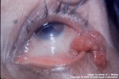
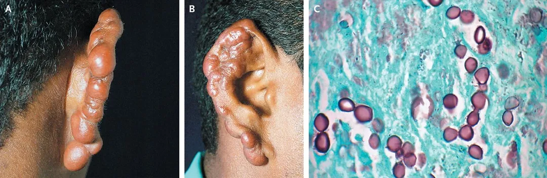
Hard, painless nodules (keloids) on the face, ears, and upper extremities are a hallmark of lobomycosis.
Lesions of mycetoma and chromoblastomycosis are comparable to those of lobstermycosis. Upon direct microscopic inspection of removed tissue in a KOH solution, lengthy diagnostic chains of yeast cells are visible.
For lobomycosis, surgical excision is curative.
Lobomycosis
Caused by Loboa loboi, lobomycosis is frequently linked to a traumatic agent injection. Dolphins naturally contract L loboi.
Hard, painless nodules (keloids) on the face, ears, and upper extremities are a hallmark of lobomycosis.
Lesions of mycetoma and chromoblastomycosis are comparable to those of lobstermycosis. Upon direct microscopic inspection of removed tissue in a KOH solution, lengthy diagnostic chains of yeast cells are visible.
For lobomycosis, surgical excision is curative.

- Published on
Infectious Diseases and Microbiology - Hantavirus Pulmonary Syndrome ( Hantavirus)
HPS, or hantavirus pulmonary syndrome
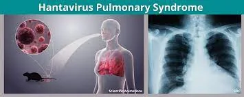
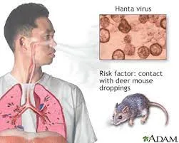
HPS is a viral zoonosis caused by the hantavirus, a member of the bunyavirus family. Aerosol inhalation of rat excrement contaminated with the hantavirus is the method of transmission. The rodent reservoir for HPS is the deer mouse. In the US, there has never been any evidence of hantavirus transmission from person to person. The most significant hantavirus in the US and Canada is the Sin Nombre virus.
A brief prodromal sickness that resembles influenza and is marked by fever, myalgia, headache, coughing, and gastrointestinal symptoms is HPS. The illness advances quickly, resulting in hypotension, pulmonary edema, thrombocytopenia, and dyspnea. For most patients, mechanical ventilation is necessary. The death rate from HPS is substantial (>50%) in previously healthy persons.
In pulmonary capillary endothelial cells, the hantavirus multiplies. Upon postmortem, HPS patients exhibited bilateral pleural effusions accompanied by mononuclear cell infiltrates in the interstitial space. One possible aspect of HPS pathogenesis is immune damage to virus-infected endothelium cells. Immunity mediated by humour and cells is thought to be in charge of recuperation and defense against recurrent infections.
Serologic testing for hantavirus IgM and IgG in serum is used to establish the diagnosis. Antigen of the hantavirus
Lung tissue can be used for diagnosis using immunohistochemistry or PCR to detect viral RNA.
Treatment and A vaccine for hantavirus infection are not available. As soon as HPS symptoms appear, immediate intensive treatment is required. Avoiding direct contact with diseased deer, mice, or their excrement is the best method of control.
HPS, or hantavirus pulmonary syndrome
HPS is a viral zoonosis caused by the hantavirus, a member of the bunyavirus family. Aerosol inhalation of rat excrement contaminated with the hantavirus is the method of transmission. The rodent reservoir for HPS is the deer mouse. In the US, there has never been any evidence of hantavirus transmission from person to person. The most significant hantavirus in the US and Canada is the Sin Nombre virus.
A brief prodromal sickness that resembles influenza and is marked by fever, myalgia, headache, coughing, and gastrointestinal symptoms is HPS. The illness advances quickly, resulting in hypotension, pulmonary edema, thrombocytopenia, and dyspnea. For most patients, mechanical ventilation is necessary. The death rate from HPS is substantial (>50%) in previously healthy persons.
In pulmonary capillary endothelial cells, the hantavirus multiplies. Upon postmortem, HPS patients exhibited bilateral pleural effusions accompanied by mononuclear cell infiltrates in the interstitial space. One possible aspect of HPS pathogenesis is immune damage to virus-infected endothelium cells. Immunity mediated by humour and cells is thought to be in charge of recuperation and defense against recurrent infections.
Serologic testing for hantavirus IgM and IgG in serum is used to establish the diagnosis. Antigen of the hantavirus
Lung tissue can be used for diagnosis using immunohistochemistry or PCR to detect viral RNA.
Treatment and A vaccine for hantavirus infection are not available. As soon as HPS symptoms appear, immediate intensive treatment is required. Avoiding direct contact with diseased deer, mice, or their excrement is the best method of control.
- Published on
Infectious Diseases and Microbiology - Lassa Fever (Lassa Fever Virus)
Lassa Fever
The Lassa fever virus, a member of the arenavirus family, is the cause of Lassa fever, a viral zoonosis. The Lassa virus is spread by direct contact with the excrement of infected rodents or by inhaling aerosolized particles. Lassa fever can also transfer between individuals by direct contact with an infected person's blood, tissue, secretions, or excretions. The rodent reservoir for infection is the field rat. West Africa is home to the endemic Lassa fever. The hospitalized patient's case mortality rate ranges from 15% to 20%.
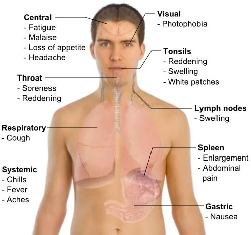
Fever, myalgia, and severe prostration are the hallmarks of severe hemorrhagic fever caused by the Lassa fever virus. CNS and hemorrhagic symptoms appear later. Typical side effects include spontaneous abortion and hearing loss.
The Lassa virus enters the body through skin abrasions or inhalation, replicates in local lymph nodes, and then causes viremia. Once inside macrophages, the virus travels throughout the body. The release of cytokines by infected macrophages is linked to mortality and adds to disease.
The usual laboratory approach for diagnosis is serologic testing for IgM or IgG specific to the Lassa virus.
Patients with severe Lassa fever respond well to therapy with ribavirin. It has been discovered that convalescent serum from individuals who have recovered from the illness can be helpful in some situations. There isn't a vaccine available. Strict infection control procedures and rodent management are crucial for prevention.
Lassa Fever
The Lassa fever virus, a member of the arenavirus family, is the cause of Lassa fever, a viral zoonosis. The Lassa virus is spread by direct contact with the excrement of infected rodents or by inhaling aerosolized particles. Lassa fever can also transfer between individuals by direct contact with an infected person's blood, tissue, secretions, or excretions. The rodent reservoir for infection is the field rat. West Africa is home to the endemic Lassa fever. The hospitalized patient's case mortality rate ranges from 15% to 20%.
Fever, myalgia, and severe prostration are the hallmarks of severe hemorrhagic fever caused by the Lassa fever virus. CNS and hemorrhagic symptoms appear later. Typical side effects include spontaneous abortion and hearing loss.
The Lassa virus enters the body through skin abrasions or inhalation, replicates in local lymph nodes, and then causes viremia. Once inside macrophages, the virus travels throughout the body. The release of cytokines by infected macrophages is linked to mortality and adds to disease.
The usual laboratory approach for diagnosis is serologic testing for IgM or IgG specific to the Lassa virus.
Patients with severe Lassa fever respond well to therapy with ribavirin. It has been discovered that convalescent serum from individuals who have recovered from the illness can be helpful in some situations. There isn't a vaccine available. Strict infection control procedures and rodent management are crucial for prevention.
Symptoms and Signs of Lassa Fever
- Published on
Infectious Diseases and Microbiology - Lymphocytic Choriomeningitis (lymphcytic Choriomeningitis Virus)
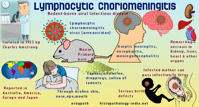
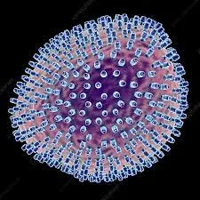
The LCM virus (LCMV), a member of the arenavirus family, is the cause of LCM, a viral zoonosis. Aerosol inhalation, eating contaminated food, or coming into contact with the secretions or excretions of infected rats are the three main ways that LCMV is spread. Except for rare cases of organ transplantation from an LCMV-infected donor and vertical transmission from an infected mother to fetus, person-to-person transmission has not been documented. The typical infection reservoirs are house mice or pet hamsters. The global dissemination of LCMV is accompanied by a low case fatality rate (<1%).
Asymptomatic infections with LCMV predominate. Malaise, headache, myalgia, fever, and other influenza-like symptoms can be signs of symptomatic infections. In a small percentage of cases, LCMV results in aseptic meningitis, which is characterized by fever, headache, and stiff neck. The majority of patients fully recover. Weakness, despair, and trouble concentrating are examples of CNS problems that might last for weeks.
Congenital hydrocephalus, chorioretinitis, spontaneous miscarriage, and mental retardation have all been linked to infections contracted during pregnancy.
After entering the body through skin abrasions, ingestion, or aerosol inhalation, LCMV replicates in lymph nodes before causing viremia. The virus propagates throughout the body by replicating in macrophages. Meningitis tissue damage is associated with cytokine release and cytotoxic T-cell-induced immunopathology.
Normal diagnosis involves serologic testing for virus-specific IgM or IgG.
For LCMV infection, there isn't a specific medication or vaccination available. Prevention requires effective rodent control.
The LCM virus (LCMV), a member of the arenavirus family, is the cause of LCM, a viral zoonosis. Aerosol inhalation, eating contaminated food, or coming into contact with the secretions or excretions of infected rats are the three main ways that LCMV is spread. Except for rare cases of organ transplantation from an LCMV-infected donor and vertical transmission from an infected mother to fetus, person-to-person transmission has not been documented. The typical infection reservoirs are house mice or pet hamsters. The global dissemination of LCMV is accompanied by a low case fatality rate (<1%).
Asymptomatic infections with LCMV predominate. Malaise, headache, myalgia, fever, and other influenza-like symptoms can be signs of symptomatic infections. In a small percentage of cases, LCMV results in aseptic meningitis, which is characterized by fever, headache, and stiff neck. The majority of patients fully recover. Weakness, despair, and trouble concentrating are examples of CNS problems that might last for weeks.
Congenital hydrocephalus, chorioretinitis, spontaneous miscarriage, and mental retardation have all been linked to infections contracted during pregnancy.
After entering the body through skin abrasions, ingestion, or aerosol inhalation, LCMV replicates in lymph nodes before causing viremia. The virus propagates throughout the body by replicating in macrophages. Meningitis tissue damage is associated with cytokine release and cytotoxic T-cell-induced immunopathology.
Normal diagnosis involves serologic testing for virus-specific IgM or IgG.
For LCMV infection, there isn't a specific medication or vaccination available. Prevention requires effective rodent control.
- Published on
Infectious Diseases and Microbiology - Gastroenteritis (Rotavirus)
Gastroenteritis caused by Rotavirus
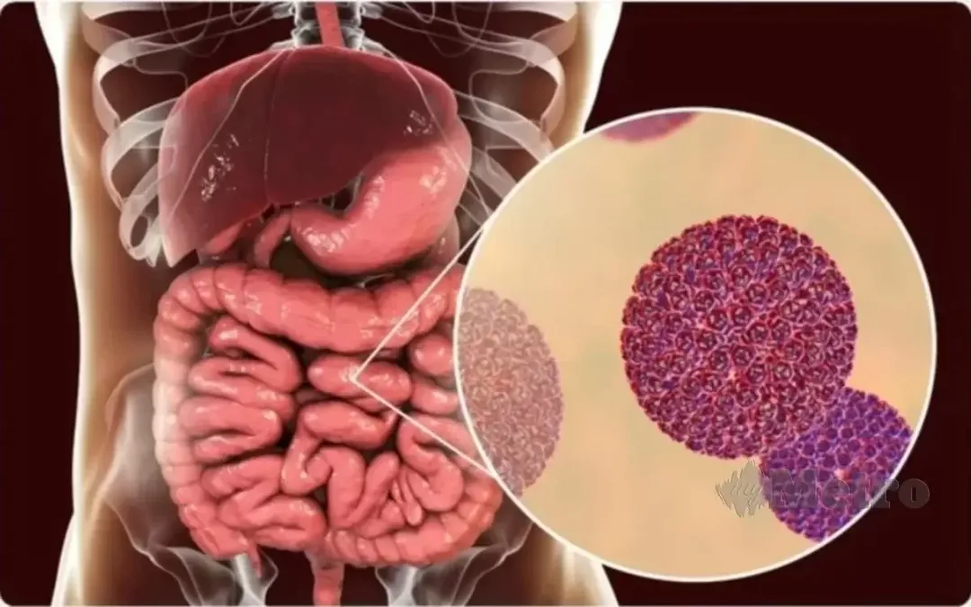
Acute viral gastroenteritis is mainly caused by rotavirus in newborns and young children. Rotavirus spreads through the fecal-oral pathway. Globally, infections are common, and severe diarrhea is thought to be the cause of one million newborn deaths, especially in developing nations. All age groups can contract rotavirus gastroenteritis, however children between the ages of 6 and 24 months have the highest rate of severe illness.
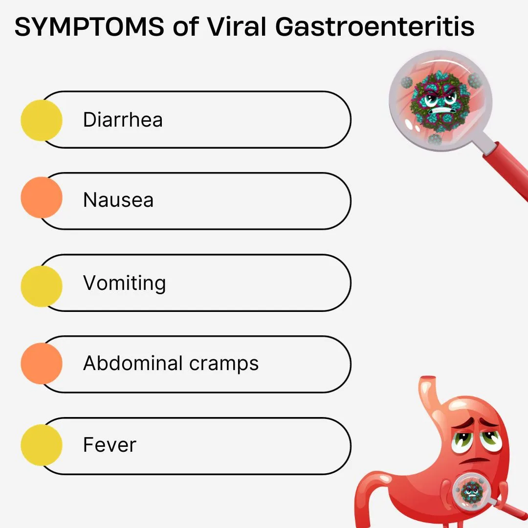
The symptoms of a rotavirus infection include vomiting, low-grade fever, nausea, and non-bloody, watery diarrhea that lasts for four to five days. The two main side effects of severe diarrhea are electrolyte loss and dehydration.
Individuals who are malnourished and have concomitant immunodeficiencies are more vulnerable to severe rotavirus infections.
Rotaviruses infect and lyse the small intestine's enterocytes, which are epithelial cells, after ingestion. Stunted villi subsequently result from damaged enterocytes sloughing off. A flow of excess intestinal fluid across the intestinal membrane is linked to damage to enterocytes, and in certain cases, this leads to a net loss of salt and protein. Damage to the mucosa reduces the small intestine's absorptive surface area and the amount of digestive enzymes (such disaccharidases) that are produced. Due to a malabsorptive state brought on by these inadequacies, there is a hyperosmotic impact that results in diarrhea. Serum antibody levels are associated with both viral clearance and subsequent immunity.
Immunity against reinfection is associated with the intestinal secretory IgA response.
The technique commonly employed for infection diagnosis is enzyme immunoassay detection of rotavirus antigen in feces.
For rotavirus infection, there is no particular treatment. In extreme situations, oral rehydration is necessary to replenish electrolytes and fluids. It is authorized to prevent rotavirus diarrhea in babies by administering an oral, live, attenuated pentavalent rotavirus vaccination. Better hygiene practices and sanitation protocols are control strategies.
Gastroenteritis caused by Rotavirus
Acute viral gastroenteritis is mainly caused by rotavirus in newborns and young children. Rotavirus spreads through the fecal-oral pathway. Globally, infections are common, and severe diarrhea is thought to be the cause of one million newborn deaths, especially in developing nations. All age groups can contract rotavirus gastroenteritis, however children between the ages of 6 and 24 months have the highest rate of severe illness.
The symptoms of a rotavirus infection include vomiting, low-grade fever, nausea, and non-bloody, watery diarrhea that lasts for four to five days. The two main side effects of severe diarrhea are electrolyte loss and dehydration.
Individuals who are malnourished and have concomitant immunodeficiencies are more vulnerable to severe rotavirus infections.
Rotaviruses infect and lyse the small intestine's enterocytes, which are epithelial cells, after ingestion. Stunted villi subsequently result from damaged enterocytes sloughing off. A flow of excess intestinal fluid across the intestinal membrane is linked to damage to enterocytes, and in certain cases, this leads to a net loss of salt and protein. Damage to the mucosa reduces the small intestine's absorptive surface area and the amount of digestive enzymes (such disaccharidases) that are produced. Due to a malabsorptive state brought on by these inadequacies, there is a hyperosmotic impact that results in diarrhea. Serum antibody levels are associated with both viral clearance and subsequent immunity.
Immunity against reinfection is associated with the intestinal secretory IgA response.
The technique commonly employed for infection diagnosis is enzyme immunoassay detection of rotavirus antigen in feces.
For rotavirus infection, there is no particular treatment. In extreme situations, oral rehydration is necessary to replenish electrolytes and fluids. It is authorized to prevent rotavirus diarrhea in babies by administering an oral, live, attenuated pentavalent rotavirus vaccination. Better hygiene practices and sanitation protocols are control strategies.
- Published on
Infectious Diseases and Microbiology - Colorado Tick Fever (Colorado Tick Fever Virus)
Colorado Tick Fever
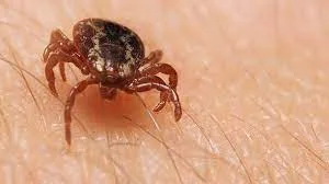
The Colorado tick fever virus (CTFV) is the cause of Colorado tick fever, a viral zoonosis. The bite of an infected Dermacentor andersoni wood tick (CTFV) spreads the arbovirus. All across the western United States, CTFV is endemic. The natural host of CTFV is small animals, such as chipmunks and squirrels.
The symptoms of CTF, an acute febrile sickness, include fever, chills, myalgia, headache, malaise, stomach discomfort, and vomiting. About half of the affected patients have a "saddle-back" fever pattern, which consists of a 2-3 day fever episode followed by an afebrile period and a subsequent fever return. Hemorrhagic fever, often known as encephalitis, is a consequence of CTFV infection, particularly in young patients.
Deaths are not common.
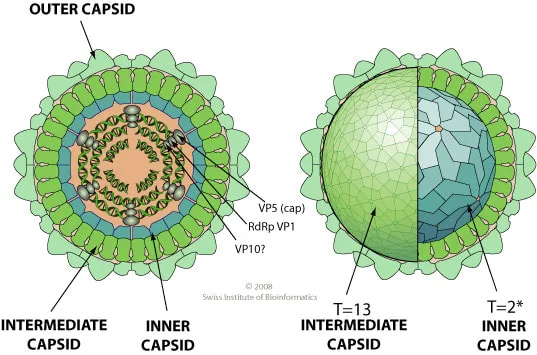
CTFV replicates in hematopoietic cells, including erythrocyte precursors, after entering the skin through a tick bite. Frequent observations of leukopenia and thrombocytopenia are likely due to direct cytopathic effects of the virus on stem cells. When mature erythrocytes are shielded from immune clearance, the virus endures. Increased neutralizing antibody levels and resistance to reinfection are linked to recovery.
Blood smears can be directly immunostaining to identify CTFV antigens on the erythrocyte surface. The diagnosis of CTFV infection can be made serologically using enzyme immunoassay to look for virus-specific IgM or IgG.
For CTFV, there isn't a particular treatment or vaccination. In tick-endemic areas, protection from ticks is the most effective control measure.
Colorado Tick Fever
The Colorado tick fever virus (CTFV) is the cause of Colorado tick fever, a viral zoonosis. The bite of an infected Dermacentor andersoni wood tick (CTFV) spreads the arbovirus. All across the western United States, CTFV is endemic. The natural host of CTFV is small animals, such as chipmunks and squirrels.
The symptoms of CTF, an acute febrile sickness, include fever, chills, myalgia, headache, malaise, stomach discomfort, and vomiting. About half of the affected patients have a "saddle-back" fever pattern, which consists of a 2-3 day fever episode followed by an afebrile period and a subsequent fever return. Hemorrhagic fever, often known as encephalitis, is a consequence of CTFV infection, particularly in young patients.
Deaths are not common.
CTFV replicates in hematopoietic cells, including erythrocyte precursors, after entering the skin through a tick bite. Frequent observations of leukopenia and thrombocytopenia are likely due to direct cytopathic effects of the virus on stem cells. When mature erythrocytes are shielded from immune clearance, the virus endures. Increased neutralizing antibody levels and resistance to reinfection are linked to recovery.
Blood smears can be directly immunostaining to identify CTFV antigens on the erythrocyte surface. The diagnosis of CTFV infection can be made serologically using enzyme immunoassay to look for virus-specific IgM or IgG.
For CTFV, there isn't a particular treatment or vaccination. In tick-endemic areas, protection from ticks is the most effective control measure.

- Published on
Infectious Diseases and Microbiology - HIV/AIDS ( HIV-1)
HIV-1 is responsible for the development of AIDS. HIV-1 is transferred by sexual intercourse, blood transfusion, intravenous medication administration, and from an infected mother to her child either through the placenta during pregnancy or during childbirth. The primary means of HIV-1 transmission in the United States is through homosexual contact. Heterosexual transmission is the predominant mode of transfer throughout the rest of the world.
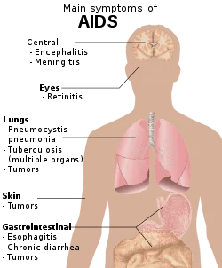
HIV disease is distinguished by an initial phase that exhibits symptoms similar to the flu or infectious mononucleosis, followed by a symptom-free phase of clinical latency marked by fatigue, weight loss, night sweats, or lymphadenopathy. On average, it takes around 10 years for AIDS to emerge during this phase. The symptomatic phase of AIDS is the final stage of the disease. AIDS is defined by a CD4+ T lymphocyte count that is lower than 200/mL (normal range is 800-1200/mL). It is also associated with opportunistic infections caused by protozoa, fungi, bacteria, and viruses, as well as the development of malignancies.
The process by which a disease develops and progresses. HIV-1 binds to the CD4 molecule and a chemokine receptor on helper T lymphocytes, monocytes-macrophages, and mucosal dendritic cells through the envelope glycoprotein gp120.
HIV-1 has the ability to infect both CD4+ cells that are active and those that are not activated in the lymph node where it is located. The virus remains dormant in nonactivated (resting) T cells, but it reproduces and destroys T cells that have been activated by infection or cytokines, or both.
The diagnosis of HIV and AIDS is made by identifying virus-specific antibodies to HIV-1 by a screening test, which is then confirmed by a Western blot assay. RT-PCR is employed to measure the quantity of HIV-1 in plasma, known as viral load, and to track the advancement of the disease and evaluate the effectiveness of antiretroviral therapy.
Three kinds of antiretroviral medicines, including nucleoside RT inhibitors, nonnucleoside RTIs, and protease inhibitors, are provided in combination for treatment. Currently, there is no available vaccination that can effectively prevent HIV-1 infection. The implementation of HIV-1 antibody screening in blood transfusions has effectively halted the transmission of HIV by this method. The transmission of HIV-1 from mother to infant can be considerably decreased with the use of antiretroviral medication for both the pregnant woman and the newborn baby.
HIV-1 is responsible for the development of AIDS. HIV-1 is transferred by sexual intercourse, blood transfusion, intravenous medication administration, and from an infected mother to her child either through the placenta during pregnancy or during childbirth. The primary means of HIV-1 transmission in the United States is through homosexual contact. Heterosexual transmission is the predominant mode of transfer throughout the rest of the world.
HIV disease is distinguished by an initial phase that exhibits symptoms similar to the flu or infectious mononucleosis, followed by a symptom-free phase of clinical latency marked by fatigue, weight loss, night sweats, or lymphadenopathy. On average, it takes around 10 years for AIDS to emerge during this phase. The symptomatic phase of AIDS is the final stage of the disease. AIDS is defined by a CD4+ T lymphocyte count that is lower than 200/mL (normal range is 800-1200/mL). It is also associated with opportunistic infections caused by protozoa, fungi, bacteria, and viruses, as well as the development of malignancies.
The process by which a disease develops and progresses. HIV-1 binds to the CD4 molecule and a chemokine receptor on helper T lymphocytes, monocytes-macrophages, and mucosal dendritic cells through the envelope glycoprotein gp120.
HIV-1 has the ability to infect both CD4+ cells that are active and those that are not activated in the lymph node where it is located. The virus remains dormant in nonactivated (resting) T cells, but it reproduces and destroys T cells that have been activated by infection or cytokines, or both.
The diagnosis of HIV and AIDS is made by identifying virus-specific antibodies to HIV-1 by a screening test, which is then confirmed by a Western blot assay. RT-PCR is employed to measure the quantity of HIV-1 in plasma, known as viral load, and to track the advancement of the disease and evaluate the effectiveness of antiretroviral therapy.
Three kinds of antiretroviral medicines, including nucleoside RT inhibitors, nonnucleoside RTIs, and protease inhibitors, are provided in combination for treatment. Currently, there is no available vaccination that can effectively prevent HIV-1 infection. The implementation of HIV-1 antibody screening in blood transfusions has effectively halted the transmission of HIV by this method. The transmission of HIV-1 from mother to infant can be considerably decreased with the use of antiretroviral medication for both the pregnant woman and the newborn baby.
- Published on
Infectious Diseases and Microbiology - Adult T- Cell Leukemia ( Human T lymphotropic virus type 1 (HTLV-1))
Human T-lymphotropic virus type 1 (HTLV-1) adult T-cell leukemia
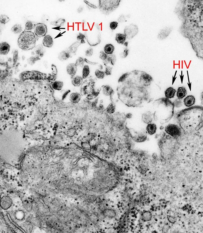
HTLV-1 is responsible for adult T-cell leukemia (ATL) and a neurological condition known as HTLV-1-associated myelopathy-tropical spastic paraparesis (HAM-TSP). HTLV-1 is predominantly linked to cells and is primarily transmitted through cells infected with HTLV-1, rather than through viral particles. HTLV-1 is spread through the transfer of infected lymphocytes present in breast milk, semen, and blood.
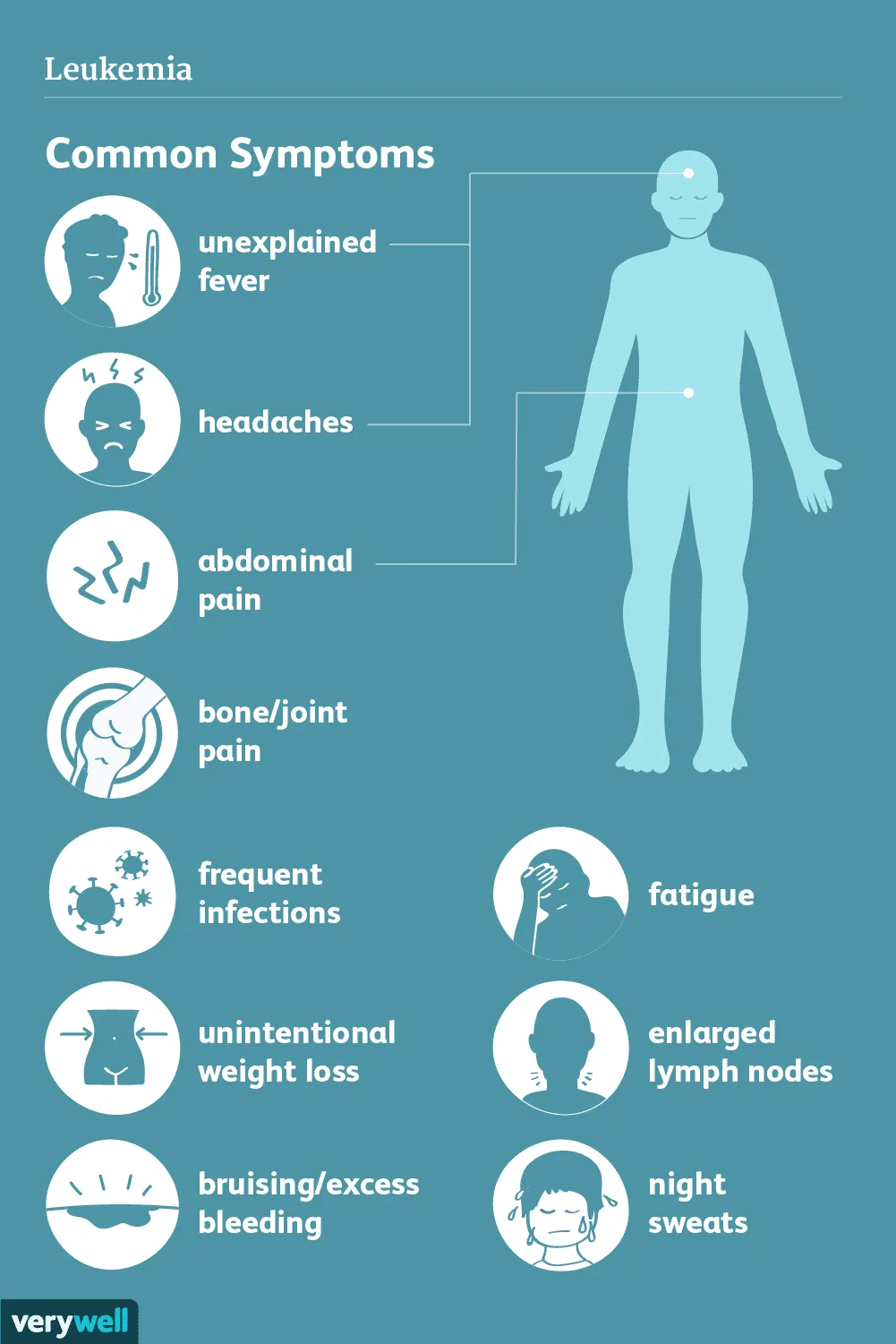
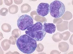
ATL is distinguished by a protracted asymptomatic phase of 20–50 years, elevated quantities of leukemia cells, skin abnormalities, widespread lymph node enlargement, enlargement of the liver and spleen, and excessive levels of calcium in the blood. The probability of an infected individual developing ATL over their lifetime is 3-5%. HAM-TSP is distinguished by a briefer amount of time before symptoms appear compared to ATL (2-4 years). It involves the damage of the long motor neurons in the spinal cord, resulting in muscle weakness in the legs, gradual stiffness, back discomfort, loss of bladder control, heightened reflexes, sensory abnormalities, and erectile dysfunction in males. The lifetime probability of developing HAM is approximately 1%.
HTLV-1 predominantly infects CD4+ T cells. The HTLV-1 encoded regulatory protein Tax enhances the proliferation and mitosis of CD4+ T lymphocytes by triggering cellular transcription factors that activate cellular growth factors and growth factor receptors, such as interleukin-2 (IL-2) and IL-2 receptor. The pathogenesis of HAM-TSP is distinguished by the invasion of HTLV-1 infected cells into the central nervous system and a significant immune response by cytotoxic lymphocytes, resulting in damage to neurological tissues.
The presence of HTLV-1 infection is identified by Enzyme Immunoassay (EIA) which detects virus-specific antibodies.
There is currently no targeted therapy or immunization available for HTLV-1. Implementing blood screening measures can effectively prevent the transmission of HTLV-1 through transfusions. Additionally, the cessation of breastfeeding by women infected with HTLV-1 will effectively eliminate the transmission of the virus from mother to newborn. Preventive methods implemented for HIV infection are also applicable for HTLV-1.
Human T-lymphotropic virus type 1 (HTLV-1) adult T-cell leukemia
HTLV-1 is responsible for adult T-cell leukemia (ATL) and a neurological condition known as HTLV-1-associated myelopathy-tropical spastic paraparesis (HAM-TSP). HTLV-1 is predominantly linked to cells and is primarily transmitted through cells infected with HTLV-1, rather than through viral particles. HTLV-1 is spread through the transfer of infected lymphocytes present in breast milk, semen, and blood.
ATL is distinguished by a protracted asymptomatic phase of 20–50 years, elevated quantities of leukemia cells, skin abnormalities, widespread lymph node enlargement, enlargement of the liver and spleen, and excessive levels of calcium in the blood. The probability of an infected individual developing ATL over their lifetime is 3-5%. HAM-TSP is distinguished by a briefer amount of time before symptoms appear compared to ATL (2-4 years). It involves the damage of the long motor neurons in the spinal cord, resulting in muscle weakness in the legs, gradual stiffness, back discomfort, loss of bladder control, heightened reflexes, sensory abnormalities, and erectile dysfunction in males. The lifetime probability of developing HAM is approximately 1%.
HTLV-1 predominantly infects CD4+ T cells. The HTLV-1 encoded regulatory protein Tax enhances the proliferation and mitosis of CD4+ T lymphocytes by triggering cellular transcription factors that activate cellular growth factors and growth factor receptors, such as interleukin-2 (IL-2) and IL-2 receptor. The pathogenesis of HAM-TSP is distinguished by the invasion of HTLV-1 infected cells into the central nervous system and a significant immune response by cytotoxic lymphocytes, resulting in damage to neurological tissues.
The presence of HTLV-1 infection is identified by Enzyme Immunoassay (EIA) which detects virus-specific antibodies.
There is currently no targeted therapy or immunization available for HTLV-1. Implementing blood screening measures can effectively prevent the transmission of HTLV-1 through transfusions. Additionally, the cessation of breastfeeding by women infected with HTLV-1 will effectively eliminate the transmission of the virus from mother to newborn. Preventive methods implemented for HIV infection are also applicable for HTLV-1.