- Published on
Infectious Diseases and Microbiology - Progressive Multifocal Leukoencephalopathy (PML) (JC Virus)
Progressive Multifocal Leukoencephalopathy (PML)
PML is a consequence of the JC virus (JCV), a polyomavirus that affects humans. JCV is transferred through respiratory droplets, primarily during early childhood, and eventually affects 70%–80% of adults. Severely immunosuppressed patients experience the reactivation of latent infections.
PML is a neurological disorder characterized by the loss of myelin in the brain, typically affecting individuals with weakened immune systems, especially those with AIDS. The beginning of the disease is gradual and difficult to detect. Initial indications manifest as atypical speech and visual impairments, along with changes in cognitive abilities. PML follows a gradual clinical trajectory, ultimately leading to a state of coma and death, typically occurring within a span of 6 months after the initial onset.
JCV is most frequently contracted through inhalation and spreads through the bloodstream to create a dormant infection in the kidneys, lungs, and lymphoid organs. Immunocompromised patients have activation of JCV, which then extends to the brain and leads to the development of PML. Within the central nervous system (CNS), the JC virus (JCV) binds to the serotonin receptor located on oligodendrocytes, resulting in a lytic infection. Oligodendrocytes are the primary cells responsible for creating myelin in the central nervous system (CNS). JCV utilizes opportunistic immune evasion mechanisms in immunosuppressed individuals.
The presence of JCV infection can be identified through the use of PCR on cerebrospinal fluid (CSF) or brain biopsy samples obtained from individuals with progressive multifocal leukoencephalopathy (PML).
There is currently no targeted therapy or immunization available for the treatment or prevention of PML.
PML is a medical condition that is used to define the presence of AIDS.
Progressive Multifocal Leukoencephalopathy (PML)
PML is a consequence of the JC virus (JCV), a polyomavirus that affects humans. JCV is transferred through respiratory droplets, primarily during early childhood, and eventually affects 70%–80% of adults. Severely immunosuppressed patients experience the reactivation of latent infections.
PML is a neurological disorder characterized by the loss of myelin in the brain, typically affecting individuals with weakened immune systems, especially those with AIDS. The beginning of the disease is gradual and difficult to detect. Initial indications manifest as atypical speech and visual impairments, along with changes in cognitive abilities. PML follows a gradual clinical trajectory, ultimately leading to a state of coma and death, typically occurring within a span of 6 months after the initial onset.
JCV is most frequently contracted through inhalation and spreads through the bloodstream to create a dormant infection in the kidneys, lungs, and lymphoid organs. Immunocompromised patients have activation of JCV, which then extends to the brain and leads to the development of PML. Within the central nervous system (CNS), the JC virus (JCV) binds to the serotonin receptor located on oligodendrocytes, resulting in a lytic infection. Oligodendrocytes are the primary cells responsible for creating myelin in the central nervous system (CNS). JCV utilizes opportunistic immune evasion mechanisms in immunosuppressed individuals.
The presence of JCV infection can be identified through the use of PCR on cerebrospinal fluid (CSF) or brain biopsy samples obtained from individuals with progressive multifocal leukoencephalopathy (PML).
There is currently no targeted therapy or immunization available for the treatment or prevention of PML.
PML is a medical condition that is used to define the presence of AIDS.


- Published on
Infectious Diseases and Microbiology - Influenza ( Influenza Virus)
Influenza A, B, and C viruses are the culprits that cause influenza. Influenza A is a contagious illness that can infect horses, birds, pigs, and people. Only humans can contract influenza B, which has a milder pandemic phenotype. Rarely, influenza C leads to subclinical illness. Respiratory droplets transmit influenza. Influenza A and B can cause antigenic drift, which involves minor changes in H or N antigens due to point mutations, or antigenic shift, which results in a major change in H or N antigens or both as a result of RNA gene segments being reassigned between the two influenza viruses. These two methods can cause antigenic variation of the influenza virus envelope proteins, hemagglutinin (H) or neuraminidase (N). In 2009, a fresh H1N1 influenza pandemic struck the world. The elderly were mainly immune to H1N1, but young people and pregnant women were particularly vulnerable.
The symptoms of influenza can appear suddenly and include fever, chills, headache, myalgia, prostration, and fever. Rhinitis and a dry cough are the next symptoms to appear. Usually taking 2-4 weeks, recovery is sluggish and accompanied by lethargy, coughing, and exhaustion.
Influenza complications might include primary viral pneumonia (uncommon) and secondary bacterial pneumonia (common). Those with chronic heart or lung diseases as well as the elderly are at risk of problems.
Cell death occurs when the influenza virus attacks respiratory epithelial cells. Recovery from infection is linked to interferon, secretory IgA specific to the virus, and cytotoxic T-cell responses.
Viral antigen can be found in clinical specimens using immunocytochemistry or enzyme immunoassay.
In cell culture, the diagnosis virus can be isolated and identified.
Amantadine and rimantadine are antiviral medications used to treat influenza A; oseltamivir and zanamivir, which are neuraminidase inhibitors, are used to treat influenza B. The best way to avoid influenza and its complications is to have an annual vaccination against strains of influenza A and B. There is a live, attenuated influenza A vaccination available for children and adults in good health.
Influenza A, B, and C viruses are the culprits that cause influenza. Influenza A is a contagious illness that can infect horses, birds, pigs, and people. Only humans can contract influenza B, which has a milder pandemic phenotype. Rarely, influenza C leads to subclinical illness. Respiratory droplets transmit influenza. Influenza A and B can cause antigenic drift, which involves minor changes in H or N antigens due to point mutations, or antigenic shift, which results in a major change in H or N antigens or both as a result of RNA gene segments being reassigned between the two influenza viruses. These two methods can cause antigenic variation of the influenza virus envelope proteins, hemagglutinin (H) or neuraminidase (N). In 2009, a fresh H1N1 influenza pandemic struck the world. The elderly were mainly immune to H1N1, but young people and pregnant women were particularly vulnerable.
The symptoms of influenza can appear suddenly and include fever, chills, headache, myalgia, prostration, and fever. Rhinitis and a dry cough are the next symptoms to appear. Usually taking 2-4 weeks, recovery is sluggish and accompanied by lethargy, coughing, and exhaustion.
Influenza complications might include primary viral pneumonia (uncommon) and secondary bacterial pneumonia (common). Those with chronic heart or lung diseases as well as the elderly are at risk of problems.
Cell death occurs when the influenza virus attacks respiratory epithelial cells. Recovery from infection is linked to interferon, secretory IgA specific to the virus, and cytotoxic T-cell responses.
Viral antigen can be found in clinical specimens using immunocytochemistry or enzyme immunoassay.
In cell culture, the diagnosis virus can be isolated and identified.
Amantadine and rimantadine are antiviral medications used to treat influenza A; oseltamivir and zanamivir, which are neuraminidase inhibitors, are used to treat influenza B. The best way to avoid influenza and its complications is to have an annual vaccination against strains of influenza A and B. There is a live, attenuated influenza A vaccination available for children and adults in good health.


- Published on
Infectious Diseases and Microbiology - Ebola Hemorrhagic Fever ( Marburg and Ebola Viruses)
Ebola Hemorrhagic Fever
Serious hemorrhagic fever is caused by filoviruses such as the Marburg and Ebola viruses. Direct contact with bodily fluids tainted with the virus causes transmission. The fatality rate from the Marburg and Ebola viruses is about 90%. It's uncertain where the viruses originate naturally.
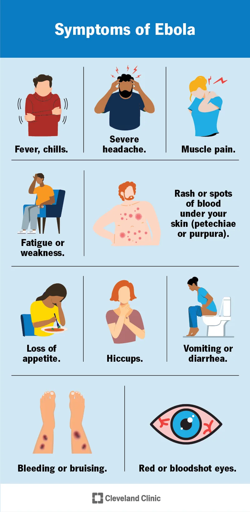
Abdominal pain, vomiting, and diarrhea are the next symptoms that accompany a quick onset of fever, headache, and joint and muscle pain, which is known as severe hemorrhagic fever. When bleeding occurs into the skin, mucous membranes, or visceral organs, the symptoms are worse. Shock and multiple organ failure cause death.
The viruses begin by infecting macrophages, then they travel throughout the body through the lymphatics and blood, infecting and necrotizing the spleen, liver, and lymph nodes. Vascular permeability, bleeding, and shock are brought on by tissue damage and elevated cytokine levels.
Viral antigens and viral RNA are detected by immunoassay and PCR, respectively, while virus-specific IgM or IgG is found by serology.
Ebola and Marburg hemorrhagic fever have no known cure or vaccine. In order to avoid direct contact with the patient and their bodily fluids, barrier measures and the isolation of suspected or confirmed infections are employed.
Ebola Hemorrhagic Fever
Serious hemorrhagic fever is caused by filoviruses such as the Marburg and Ebola viruses. Direct contact with bodily fluids tainted with the virus causes transmission. The fatality rate from the Marburg and Ebola viruses is about 90%. It's uncertain where the viruses originate naturally.
Abdominal pain, vomiting, and diarrhea are the next symptoms that accompany a quick onset of fever, headache, and joint and muscle pain, which is known as severe hemorrhagic fever. When bleeding occurs into the skin, mucous membranes, or visceral organs, the symptoms are worse. Shock and multiple organ failure cause death.
The viruses begin by infecting macrophages, then they travel throughout the body through the lymphatics and blood, infecting and necrotizing the spleen, liver, and lymph nodes. Vascular permeability, bleeding, and shock are brought on by tissue damage and elevated cytokine levels.
Viral antigens and viral RNA are detected by immunoassay and PCR, respectively, while virus-specific IgM or IgG is found by serology.
Ebola and Marburg hemorrhagic fever have no known cure or vaccine. In order to avoid direct contact with the patient and their bodily fluids, barrier measures and the isolation of suspected or confirmed infections are employed.
- Published on
Infectious Diseases and Microbiology - La Crosse Encephalitis ( La Crosse Virus)
La Crosse Encephalitis
The California encephalitis (CE) virus serogroup's subtype, La Crosse virus, is a member of the bunyavirus family and the cause of La Crosse encephalitis. The La Crosse/CE virus is an arbovirus that spreads by mosquito bites. Youngsters under 16 are most vulnerable to LaCrosse/CE. The most often reported arbovirus encephalitis in the US is La Crosse/CE, which is prevalent in the Midwest.
The majority of La Crosse/CE virus infections are asymptomatic or cause a minor case of fever.
The La Crosse/CE virus causes encephalitis, which is characterized by an abrupt onset of fever, headache, malaise, nausea, and vomiting. About 50% of encephalitis patients experience seizures. With a case fatality rate of roughly 1%, the illness normally goes away in 5-7 days. For certain patients, seizure disorders may be a consequence.
Pathophysiology A viremia is caused by an infected mosquito biting you. Transmission of the virus to CNS target tissue is made possible by the development of a secondary viremia. Antiviral antibodies offer protection against reinfection and are crucial for the removal of viruses and the resolution of infections.
Serologic testing for IgM and IgG antibodies to the La Crosse/CE Diagnosis virus is typically used to establish the diagnosis.
The La Crosse/CE virus does not have a specific therapy or vaccine. Insecticides have a significant role in controlling mosquito vectors.
La Crosse Encephalitis
The California encephalitis (CE) virus serogroup's subtype, La Crosse virus, is a member of the bunyavirus family and the cause of La Crosse encephalitis. The La Crosse/CE virus is an arbovirus that spreads by mosquito bites. Youngsters under 16 are most vulnerable to LaCrosse/CE. The most often reported arbovirus encephalitis in the US is La Crosse/CE, which is prevalent in the Midwest.
The majority of La Crosse/CE virus infections are asymptomatic or cause a minor case of fever.
The La Crosse/CE virus causes encephalitis, which is characterized by an abrupt onset of fever, headache, malaise, nausea, and vomiting. About 50% of encephalitis patients experience seizures. With a case fatality rate of roughly 1%, the illness normally goes away in 5-7 days. For certain patients, seizure disorders may be a consequence.
Pathophysiology A viremia is caused by an infected mosquito biting you. Transmission of the virus to CNS target tissue is made possible by the development of a secondary viremia. Antiviral antibodies offer protection against reinfection and are crucial for the removal of viruses and the resolution of infections.
Serologic testing for IgM and IgG antibodies to the La Crosse/CE Diagnosis virus is typically used to establish the diagnosis.
The La Crosse/CE virus does not have a specific therapy or vaccine. Insecticides have a significant role in controlling mosquito vectors.


- Published on
Infectious Diseases and Microbiology - SARS, or severe acute respiratory syndrome ( Coronavirus ( Cov))
It is a new coronavirus (CoV) that causes SARS. Approximately 30% of cold cases are caused by coronaviruses that were previously identified. SARS CoV is spread by respiratory droplets that are exhaled or coughed, and it needs close contact between individuals to spread. Additionally, the fecal-oral pathway and contaminated footpaths might transmit infection. Hong Kong published the first report of SARS in 2003. The SARS outbreak caused more than 8,000 illnesses and almost 800 deaths globally within months of spreading to North America, South America, Europe, and Asia. There were no SARS-related fatalities in the US; instead, all cases were linked to visitors coming back from regions where SARS had been reported.
Emerging from an animal reservoir, SARS is an infectious disease that may have spread to humans.
The hallmark of SARS is a two-to seven-day incubation period, which is followed by a high fever. In 10% to 20% of cases, symptoms also include headache, body aches, and diarrhea.
After two to seven days, patients with SARS develop pneumonia, hypoxia necessitating mechanical ventilation, and a dry, productive cough. The death rate from SARS is 10%.
The full pathophysiology of SARS-CoV is unknown. Common hematologic symptoms include lymphopenia and thrombocytopenia, and histology reveals diffuse alveolar destruction.
lab RT-PCR, serologic assays for the detection of CoV-specific antibodies, and viral diagnosis isolation in cell culture are the three methods used to identify SARS CoV.
There isn't a specific SARS CoV vaccination or treatment. Isolating sick people and quarantining those who have been exposed to SARS CoV are preventive methods to stop the spread of the virus. Frequent hand washing, travel restrictions to SARS-affected locations, and the use of personal protective equipment by healthcare professionals are among the steps used to stop the transmission and exposure of SARS CoV.
It is a new coronavirus (CoV) that causes SARS. Approximately 30% of cold cases are caused by coronaviruses that were previously identified. SARS CoV is spread by respiratory droplets that are exhaled or coughed, and it needs close contact between individuals to spread. Additionally, the fecal-oral pathway and contaminated footpaths might transmit infection. Hong Kong published the first report of SARS in 2003. The SARS outbreak caused more than 8,000 illnesses and almost 800 deaths globally within months of spreading to North America, South America, Europe, and Asia. There were no SARS-related fatalities in the US; instead, all cases were linked to visitors coming back from regions where SARS had been reported.
Emerging from an animal reservoir, SARS is an infectious disease that may have spread to humans.
The hallmark of SARS is a two-to seven-day incubation period, which is followed by a high fever. In 10% to 20% of cases, symptoms also include headache, body aches, and diarrhea.
After two to seven days, patients with SARS develop pneumonia, hypoxia necessitating mechanical ventilation, and a dry, productive cough. The death rate from SARS is 10%.
The full pathophysiology of SARS-CoV is unknown. Common hematologic symptoms include lymphopenia and thrombocytopenia, and histology reveals diffuse alveolar destruction.
lab RT-PCR, serologic assays for the detection of CoV-specific antibodies, and viral diagnosis isolation in cell culture are the three methods used to identify SARS CoV.
There isn't a specific SARS CoV vaccination or treatment. Isolating sick people and quarantining those who have been exposed to SARS CoV are preventive methods to stop the spread of the virus. Frequent hand washing, travel restrictions to SARS-affected locations, and the use of personal protective equipment by healthcare professionals are among the steps used to stop the transmission and exposure of SARS CoV.

- Published on
Infectious Diseases and Microbiology - Croup ( Parainfluenza viruses type 1 and 2)
The most common cause of croup (laryngotracheobronchitis) in newborns and early children is parainfluenza viruses (PIV) types 1 and 2. PIV is spread through direct contact with secretions or fomites, as well as breathing droplets. Immunity is ephemeral and reinfections frequent.
PIV 1–4 comprises the four recognized serotypes. Indicators of croup (PIV 1 and 2) include fever, barking cough, hoarseness, and inspiratory stridor. PIV 3 is linked to pneumonia and bronchiolitis in young children and babies. In both adults and children, PIV 4 causes a moderate upper respiratory infection.
PIV damages and infects respiratory epithelial cells without spreading throughout the body. The appearance of multinucleated giant cells, a virulence factor that allows direct cell-to-cell dissemination and encourages evasion from host antibodies, is one of the histopathologic modifications.
PIV diagnosis can be made quickly and directly by looking for the viral antigen in nasal aspirate cells. Alternatively, the virus can be isolated in cell culture and identified using PIV-specific antibodies.
PIV does not currently have a specific treatment or vaccination.
The most common cause of croup (laryngotracheobronchitis) in newborns and early children is parainfluenza viruses (PIV) types 1 and 2. PIV is spread through direct contact with secretions or fomites, as well as breathing droplets. Immunity is ephemeral and reinfections frequent.
PIV 1–4 comprises the four recognized serotypes. Indicators of croup (PIV 1 and 2) include fever, barking cough, hoarseness, and inspiratory stridor. PIV 3 is linked to pneumonia and bronchiolitis in young children and babies. In both adults and children, PIV 4 causes a moderate upper respiratory infection.
PIV damages and infects respiratory epithelial cells without spreading throughout the body. The appearance of multinucleated giant cells, a virulence factor that allows direct cell-to-cell dissemination and encourages evasion from host antibodies, is one of the histopathologic modifications.
PIV diagnosis can be made quickly and directly by looking for the viral antigen in nasal aspirate cells. Alternatively, the virus can be isolated in cell culture and identified using PIV-specific antibodies.
PIV does not currently have a specific treatment or vaccination.


- Published on
Infectious Diseases and Microbiology - Human Metapneumovirus Bronchiolitis (HMPV) ( Human Metapneumovirus )
As a member of the paramyxovirus family, HMPV causes pneumonia and bronchiolitis in neonates.
HMPV, which was identified in 2001, exhibits a global distribution. Although the mode of transmission remains uncertain, it is likely through respiratory droplets. Similar to RSV and influenza, HMPV infections exhibit a seasonal pattern characterized by recurrent epidemics in the winter.
The clinical manifestations of HMPV infection in neonates and children resemble those of RSV disease, which includes pneumonia, bronchiolitis, dyspnea, rhinorrhea, and wheezing.
Infections caused by HMPV are typically milder than those caused by RSV. Otitis media is a condition that is linked to HMPV infections in minors. Individuals who are immunocompromised or elderly are also susceptible to developing mild to severe HMPV infections. Mild respiratory tract infections may be caused by HMPV in the general population.
HMPV targets the respiratory tract and infects respiratory epithelial cells. Although not fully comprehended, the pathogenesis of HMPV infection is believed to be similar to that of RSV infection.
Aseptic clinical specimens are subjected to PCR analysis in order to diagnose HMPV infections.
Diagnosis Treatment and HMPV infection lacks a specific treatment or vaccine.
As a member of the paramyxovirus family, HMPV causes pneumonia and bronchiolitis in neonates.
HMPV, which was identified in 2001, exhibits a global distribution. Although the mode of transmission remains uncertain, it is likely through respiratory droplets. Similar to RSV and influenza, HMPV infections exhibit a seasonal pattern characterized by recurrent epidemics in the winter.
The clinical manifestations of HMPV infection in neonates and children resemble those of RSV disease, which includes pneumonia, bronchiolitis, dyspnea, rhinorrhea, and wheezing.
Infections caused by HMPV are typically milder than those caused by RSV. Otitis media is a condition that is linked to HMPV infections in minors. Individuals who are immunocompromised or elderly are also susceptible to developing mild to severe HMPV infections. Mild respiratory tract infections may be caused by HMPV in the general population.
HMPV targets the respiratory tract and infects respiratory epithelial cells. Although not fully comprehended, the pathogenesis of HMPV infection is believed to be similar to that of RSV infection.
Aseptic clinical specimens are subjected to PCR analysis in order to diagnose HMPV infections.
Diagnosis Treatment and HMPV infection lacks a specific treatment or vaccine.


- Published on

Infectious Diseases and Microbiology - Measles ( Measles virus)
The measles virus, a paramyxovirus with a single serotype, is the cause of the disease. Droplets in the air are the mode of transmission. With an infection rate of 85% to 95%, the measles virus is extremely contagious.
Prodromal symptoms of measles include fever, cough, coryza, and conjunctivitis. After one to two days, the cheeks develop Koplik spots, which are little white patches on irritated buccal mucosa. A day later, the head develops a maculopapular rash that lasts for three to five days and spreads to the trunk and extremities.
Measles complications include encephalitis, virus-induced giant-cell pneumonia, opportunistic bacterial superinfections (otitis media, pneumonia), and subacute sclerosing panencephalitis, a rare late-progressive neurologic disease that develops months or years after clinical measles. These complications are more common in developing nations, in malnourished children, and in immunocompromised people.
The measles virus first infects respiratory cells before proliferating and spreading in lymph nodes and spreading by viremia to other locations, such as the skin and mucosa. A cell-mediated immune response to virus-infected vascular endothelial cells in the skin causes the maculopapular rash. T and B cell infection is the primary cause of secondary infections that lead to morbidity and mortality, as well as a weakened immune response. A cell-mediated immune response is necessary for the measles to heal.
The most widely used laboratory method for measles virus-specific antibody detection is serologic testing.
Assessment, Management, and The measles virus infection has no particular therapy. The vaccination against the live, attenuated measles virus is quite successful in avoiding the disease. The vaccine is often administered in conjunction with the MMR (measles, rabies, and rubella) immunizations.
The measles virus, a paramyxovirus with a single serotype, is the cause of the disease. Droplets in the air are the mode of transmission. With an infection rate of 85% to 95%, the measles virus is extremely contagious.
Prodromal symptoms of measles include fever, cough, coryza, and conjunctivitis. After one to two days, the cheeks develop Koplik spots, which are little white patches on irritated buccal mucosa. A day later, the head develops a maculopapular rash that lasts for three to five days and spreads to the trunk and extremities.
Measles complications include encephalitis, virus-induced giant-cell pneumonia, opportunistic bacterial superinfections (otitis media, pneumonia), and subacute sclerosing panencephalitis, a rare late-progressive neurologic disease that develops months or years after clinical measles. These complications are more common in developing nations, in malnourished children, and in immunocompromised people.
The measles virus first infects respiratory cells before proliferating and spreading in lymph nodes and spreading by viremia to other locations, such as the skin and mucosa. A cell-mediated immune response to virus-infected vascular endothelial cells in the skin causes the maculopapular rash. T and B cell infection is the primary cause of secondary infections that lead to morbidity and mortality, as well as a weakened immune response. A cell-mediated immune response is necessary for the measles to heal.
The most widely used laboratory method for measles virus-specific antibody detection is serologic testing.
Assessment, Management, and The measles virus infection has no particular therapy. The vaccination against the live, attenuated measles virus is quite successful in avoiding the disease. The vaccine is often administered in conjunction with the MMR (measles, rabies, and rubella) immunizations.


- Published on

Infectious Diseases and Microbiology -Mumps ( Mumps Virus)
The mumps virus, a paramyxovirus with a single serotype, is the cause of mumps. Urine, salivary secretions, or respiratory droplets can all spread the highly contagious mumps virus. Although school-age children frequently contract the mumps, the disease is infrequent in the US due to an efficient vaccine.
The prodromal phase of the mumps is marked by fever, malaise, and headaches, and is followed by
Parotitis symptoms include swelling, inflammation, and pain in the parotid gland. The most common mumps presentation is aseptic meningitis; encephalitis is less prevalent. Orchitis, or enlargement of the testicles in adult males, is one of the complications of the mumps. It is rare to be sterile.
Viralemia transmits the mumps virus to the salivary glands, central nervous system, and other organs when it infects respiratory epithelial cells and local lymph nodes. Edema, lymphocyte infiltration, and inflammation are the causes of parathyroid gland swelling. Urine virus infection, or viruria, is frequent. Cell-mediated immunity is necessary for the body to heal from illness.
The most popular diagnostic method is serologic identification of an antibody specific to the mumps virus.
The mumps virus infection has no particular therapy. A extremely efficient method of avoiding the mumps is vaccination with an attenuated live virus. Usually, the vaccination is administered in addition to the MMR (measles, rubella) vaccine.
The mumps virus, a paramyxovirus with a single serotype, is the cause of mumps. Urine, salivary secretions, or respiratory droplets can all spread the highly contagious mumps virus. Although school-age children frequently contract the mumps, the disease is infrequent in the US due to an efficient vaccine.
The prodromal phase of the mumps is marked by fever, malaise, and headaches, and is followed by
Parotitis symptoms include swelling, inflammation, and pain in the parotid gland. The most common mumps presentation is aseptic meningitis; encephalitis is less prevalent. Orchitis, or enlargement of the testicles in adult males, is one of the complications of the mumps. It is rare to be sterile.
Viralemia transmits the mumps virus to the salivary glands, central nervous system, and other organs when it infects respiratory epithelial cells and local lymph nodes. Edema, lymphocyte infiltration, and inflammation are the causes of parathyroid gland swelling. Urine virus infection, or viruria, is frequent. Cell-mediated immunity is necessary for the body to heal from illness.
The most popular diagnostic method is serologic identification of an antibody specific to the mumps virus.
The mumps virus infection has no particular therapy. A extremely efficient method of avoiding the mumps is vaccination with an attenuated live virus. Usually, the vaccination is administered in addition to the MMR (measles, rubella) vaccine.


- Published on

Infectious Diseases and Microbiology - Rabies ( Rabies Virus)
The rabies virus, a member of the rhabdovirus family, is the cause of rabies, a viral zoonotic infection. Aerosol contact with mucosal membranes, infectious saliva from a rabid animal bite, or (very rarely) human-to-human transmission to recipients of solid organ transplants from a rabies-infected donor are the three ways that the rabies virus can spread. In the US, foxes, coyotes, skunks, bats, and raccoons are among the natural carriers of infection.
The time it takes for rabies to incubate varies, often taking 30 to 60 days following a bite. Fever, lethargy, nausea, vomiting, and pain or itching at the bite wound site are the hallmarks of a prodromal phase. The symptoms of the neurologic phase include encephalitis, coma, hydrophobia, agitation, and hyperactivity. Rabies usually results in death.
The rabies virus grows at the bite site and attaches itself to the acetylcholine receptor to infect sensory neurons. In the central nervous system, the virus replicates in gray matter by ascending axons. The virus then travels to the skin and salivary glands by peripheral nerves. To prevent disease, cell-mediated immunity is insufficient.
The diagnosis of rabies is made either by immunochemically detecting viral antigen in autopsied brain tissue or by cytologic identification of Negri bodies (cytoplasmic eosinophilic inclusion-bodies) in neurons. Immunocytochemistry can identify rabies virus antigens in biopsy samples taken from the skin at the nape of the neck or from corneal scrapings in live patients. By using PCR, rabies virus RNA in saliva can be found.
Once clinical signs have appeared, there is no particular treatment for rabies. Prophylactic measures taken both before and after exposure help prevent rabies. Post-exposure prophylaxis, or treatment following exposure to a rabid animal bite, entails three steps: (1) a complete cleaning of the site; (2) a human rabies immune globulin injection into the wound; and (3) an active rabies vaccine injection. For people who are at a high risk of contracting rabies, such as veterinarians and animal handlers, it is advised to vaccinate against the disease in advance of exposure.
The rabies virus, a member of the rhabdovirus family, is the cause of rabies, a viral zoonotic infection. Aerosol contact with mucosal membranes, infectious saliva from a rabid animal bite, or (very rarely) human-to-human transmission to recipients of solid organ transplants from a rabies-infected donor are the three ways that the rabies virus can spread. In the US, foxes, coyotes, skunks, bats, and raccoons are among the natural carriers of infection.
The time it takes for rabies to incubate varies, often taking 30 to 60 days following a bite. Fever, lethargy, nausea, vomiting, and pain or itching at the bite wound site are the hallmarks of a prodromal phase. The symptoms of the neurologic phase include encephalitis, coma, hydrophobia, agitation, and hyperactivity. Rabies usually results in death.
The rabies virus grows at the bite site and attaches itself to the acetylcholine receptor to infect sensory neurons. In the central nervous system, the virus replicates in gray matter by ascending axons. The virus then travels to the skin and salivary glands by peripheral nerves. To prevent disease, cell-mediated immunity is insufficient.
The diagnosis of rabies is made either by immunochemically detecting viral antigen in autopsied brain tissue or by cytologic identification of Negri bodies (cytoplasmic eosinophilic inclusion-bodies) in neurons. Immunocytochemistry can identify rabies virus antigens in biopsy samples taken from the skin at the nape of the neck or from corneal scrapings in live patients. By using PCR, rabies virus RNA in saliva can be found.
Once clinical signs have appeared, there is no particular treatment for rabies. Prophylactic measures taken both before and after exposure help prevent rabies. Post-exposure prophylaxis, or treatment following exposure to a rabid animal bite, entails three steps: (1) a complete cleaning of the site; (2) a human rabies immune globulin injection into the wound; and (3) an active rabies vaccine injection. For people who are at a high risk of contracting rabies, such as veterinarians and animal handlers, it is advised to vaccinate against the disease in advance of exposure.


