- Published on
Infectious Diseases and Microbiology - Amebic dysentery ( Entamoeba histolytica)
Entamoeba histolytica is a type of single-celled parasite.
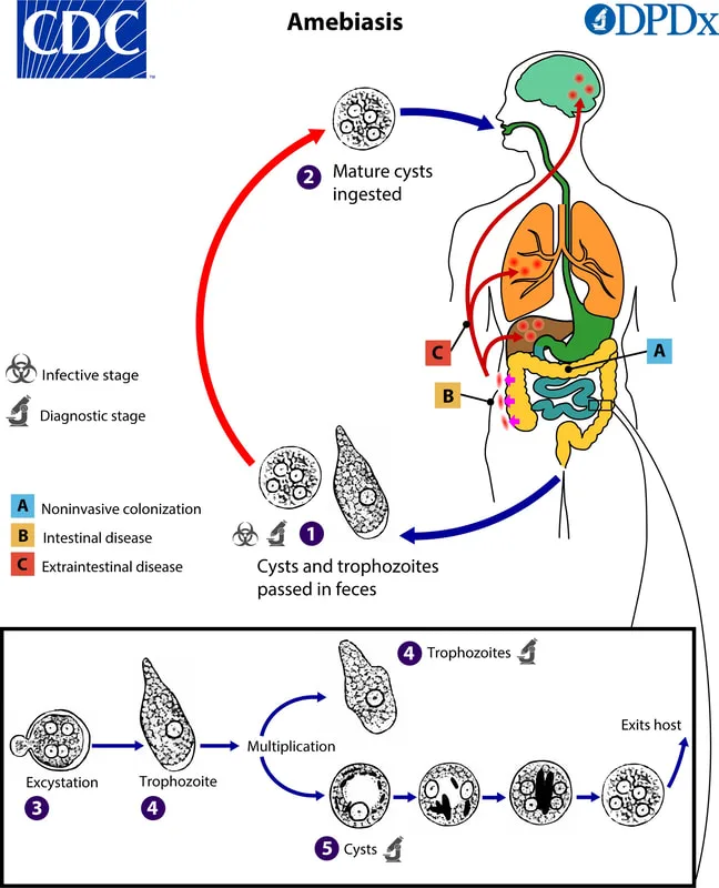
There are two stages that are encountered. The trophozoite type of amebae is the exclusive presence in tissue. It is also present in liquid feces with the occurrence of amebic dysentery. The dormant cysts are exclusively found within the colon's lumen and can be observed in soft or solid feces. Both symptomatic and asymptomatic patients excrete cysts in their feces, which can subsequently contaminate food and water sources. The cysts exhibit resistance to chlorination. The primary modes of transmission are by the ingestion of fecal matter and the direct contact of the mouth with the anus.
Infection with E histolytica leads to three distinct symptoms. Several patients are asymptomatic yet continue to be carriers, releasing cysts in their stool. patients who show symptoms may display amebic dysentery, which includes cramps, tenesmus, gas, and feces containing blood and mucus. A tiny portion of these patients may also experience more severe extraintestinal disease, characterized by abscesses, particularly in the liver.
Following the consumption of a cyst (metacyst), the process of excystation commences upon activation. The metacyst undergoes fast division, resulting in the formation of four amebulae, with each amebula corresponding to one cyst nucleus. Subsequently, each amebula undergoes further division, leading to the production of eight tiny trophozoites within each infective cyst. The trophozoites invade the large intestine, resulting in tissue damage and the formation of a distinct flask-shaped ulcer. Further invasion leads to the development of abscesses and the spread of the infection throughout the body.
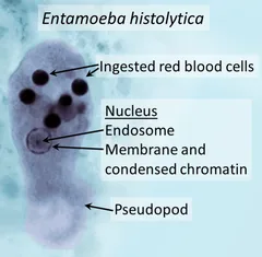
The stool analysis under a microscope shows trophozoites with ingested red blood cells and cysts with four nuclei, indicating the presence of the parasite. Nonpathogenic species, such as Entamoeba coli, can be differentiated from E histolytica by the presence of cysts that contain eight nuclei. Serum antibody testing can aid in diagnosing amebic dysentery and extraintestinal amebiasis that affects the liver.
Treatment for symptoms of varying severity may involve the administration of metronidazole and luminal amebicides, such as iodoquinol. Control strategies include the implementation of proper hygiene practices, sanitation measures, and educational initiatives aimed at preventing the transmission of diseases.
Entamoeba histolytica is a type of single-celled parasite.
There are two stages that are encountered. The trophozoite type of amebae is the exclusive presence in tissue. It is also present in liquid feces with the occurrence of amebic dysentery. The dormant cysts are exclusively found within the colon's lumen and can be observed in soft or solid feces. Both symptomatic and asymptomatic patients excrete cysts in their feces, which can subsequently contaminate food and water sources. The cysts exhibit resistance to chlorination. The primary modes of transmission are by the ingestion of fecal matter and the direct contact of the mouth with the anus.
Infection with E histolytica leads to three distinct symptoms. Several patients are asymptomatic yet continue to be carriers, releasing cysts in their stool. patients who show symptoms may display amebic dysentery, which includes cramps, tenesmus, gas, and feces containing blood and mucus. A tiny portion of these patients may also experience more severe extraintestinal disease, characterized by abscesses, particularly in the liver.
Following the consumption of a cyst (metacyst), the process of excystation commences upon activation. The metacyst undergoes fast division, resulting in the formation of four amebulae, with each amebula corresponding to one cyst nucleus. Subsequently, each amebula undergoes further division, leading to the production of eight tiny trophozoites within each infective cyst. The trophozoites invade the large intestine, resulting in tissue damage and the formation of a distinct flask-shaped ulcer. Further invasion leads to the development of abscesses and the spread of the infection throughout the body.
The stool analysis under a microscope shows trophozoites with ingested red blood cells and cysts with four nuclei, indicating the presence of the parasite. Nonpathogenic species, such as Entamoeba coli, can be differentiated from E histolytica by the presence of cysts that contain eight nuclei. Serum antibody testing can aid in diagnosing amebic dysentery and extraintestinal amebiasis that affects the liver.
Treatment for symptoms of varying severity may involve the administration of metronidazole and luminal amebicides, such as iodoquinol. Control strategies include the implementation of proper hygiene practices, sanitation measures, and educational initiatives aimed at preventing the transmission of diseases.

- Published on
Infectious Diseases and Microbiology - Histoplasmosis (Histoplasma capsulatum)
The disease histoplasmosis
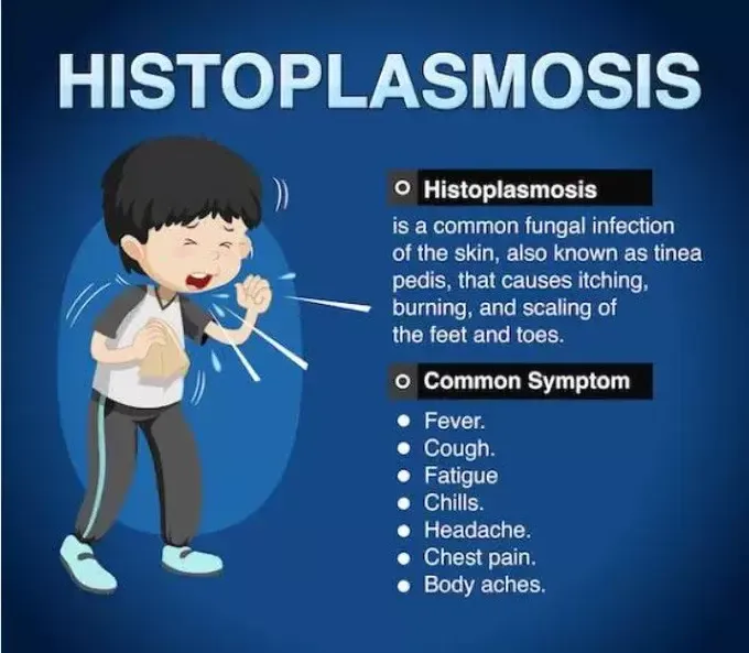
Histoplasma capsulatum is the causative agent of histoplasmosis, which is inhaled after contaminated soil is disturbed and aerosolized spores (microconidia) are released into the air. Although histoplasmosis is a global disease, it is endemic in the US in the Ohio and Mississippi River valleys. H capsulatum grows on soil, especially in soil that has been exposed to bat and bird droppings, especially those of starlings.
More than 90% of infections are self-limiting, asymptomatic, and affect immunocompetent people.
In 1% of instances, there is chronic pulmonary and progressive histoplasmosis. This condition mainly affects immunocompromised individuals or results from exposure to a high organism inoculum. Although it is uncommon, solid organ transplant recipients and immunocompromised individuals with T-cell defects—such as those with AIDS—may develop disseminated histoplasmosis. In individuals with HIV or AIDS, the death rate from disseminated histoplasmosis is approximately 10%.
Pathophysiology When H capsulatum is inhaled, the microconidia develop intracellularly like yeast grows in macrophages. This process continues until the immune system reacts, causing a localized granuloma.
A component of disseminated histoplasmosis is the invasion of reticuloendothelial system cells. H capsulatum evades phagolysosomal death by raising the pH of lysosomes, which in turn deactivates the enzymes responsible for degradation.
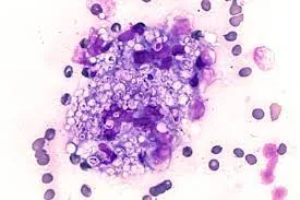
lab Yeast is visible within macrophages when clinical specimens (tissue, sputum) are examined under a microscope and stained with Wright or Giemsa stain. Patients with disseminated illness can have their blood and urine tested for a H capsulatum-specific polysaccharide antigen using an enzyme immunoassay technique.
For initial infections, which are typically self-limited, there is no need for therapy. Amphotericin B and itraconazole are used to treat histoplasmosis. A preventive method is to stay out of high endemic areas.
The disease histoplasmosis
Histoplasma capsulatum is the causative agent of histoplasmosis, which is inhaled after contaminated soil is disturbed and aerosolized spores (microconidia) are released into the air. Although histoplasmosis is a global disease, it is endemic in the US in the Ohio and Mississippi River valleys. H capsulatum grows on soil, especially in soil that has been exposed to bat and bird droppings, especially those of starlings.
More than 90% of infections are self-limiting, asymptomatic, and affect immunocompetent people.
In 1% of instances, there is chronic pulmonary and progressive histoplasmosis. This condition mainly affects immunocompromised individuals or results from exposure to a high organism inoculum. Although it is uncommon, solid organ transplant recipients and immunocompromised individuals with T-cell defects—such as those with AIDS—may develop disseminated histoplasmosis. In individuals with HIV or AIDS, the death rate from disseminated histoplasmosis is approximately 10%.
Pathophysiology When H capsulatum is inhaled, the microconidia develop intracellularly like yeast grows in macrophages. This process continues until the immune system reacts, causing a localized granuloma.
A component of disseminated histoplasmosis is the invasion of reticuloendothelial system cells. H capsulatum evades phagolysosomal death by raising the pH of lysosomes, which in turn deactivates the enzymes responsible for degradation.
lab Yeast is visible within macrophages when clinical specimens (tissue, sputum) are examined under a microscope and stained with Wright or Giemsa stain. Patients with disseminated illness can have their blood and urine tested for a H capsulatum-specific polysaccharide antigen using an enzyme immunoassay technique.
For initial infections, which are typically self-limited, there is no need for therapy. Amphotericin B and itraconazole are used to treat histoplasmosis. A preventive method is to stay out of high endemic areas.
- Published on
Infectious Diseases and Microbiology = Subacute Sclerosing Panencephalitis (SSPE)( defective measles virus mutant)
A defective measles virus mutant is the cause of SSPE. Measles virus is transmitted by respiratory droplets.
SSPE is a rare, late progressive neurologic disease of children, with the majority of cases appearing 6–8 years after acute measles. Incidence of SSPE in unvaccinated children is 1 in 1 million cases.
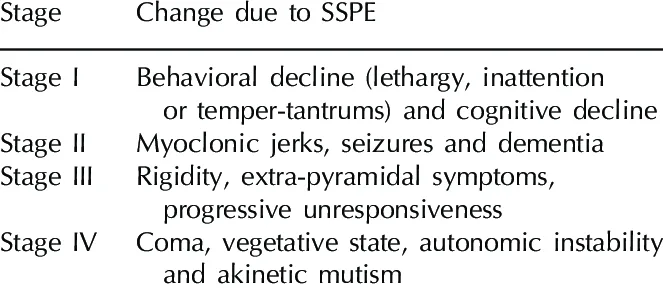
SSPE is characterized by an insidious onset of personality changes, intellectual deterioration, with later myoclonic jerks (periodic muscle spasms), spasticity, blindness, and death. The clinical course is progressive,
with death occurring 1–3 years after onset.
Brain pathology in SSPE consists of demyelination and inflammation due to chronic infection by SSPE measles virus over the course of years. Measles virus defective in virion production can be isolated from brain cells of patients with SSPE. The measles virus variant that causes SSPE has mutations in the matrix (M) protein responsible for viral assembly and budding. The lack of a functional M protein results in nonproductive infection by SSPE measles virus. Patients with SSPE have elevated levels of measles virus antibodies in serum and CSF, but no antibody to the M protein. Measles virus escapes immune surveillance by cell-to-cell fusion mediated by the fusion (F) protein.
Patients with SSPE are diagnosed by the detection of high measles virus antibody levels in serum and CSF.
There is no specific treatment for measles virus infection. Live, attenuated measles virus vaccine is highly effective in preventing measles and has reduced markedly the incidence of SSPE.
A defective measles virus mutant is the cause of SSPE. Measles virus is transmitted by respiratory droplets.
SSPE is a rare, late progressive neurologic disease of children, with the majority of cases appearing 6–8 years after acute measles. Incidence of SSPE in unvaccinated children is 1 in 1 million cases.
SSPE is characterized by an insidious onset of personality changes, intellectual deterioration, with later myoclonic jerks (periodic muscle spasms), spasticity, blindness, and death. The clinical course is progressive,
with death occurring 1–3 years after onset.
Brain pathology in SSPE consists of demyelination and inflammation due to chronic infection by SSPE measles virus over the course of years. Measles virus defective in virion production can be isolated from brain cells of patients with SSPE. The measles virus variant that causes SSPE has mutations in the matrix (M) protein responsible for viral assembly and budding. The lack of a functional M protein results in nonproductive infection by SSPE measles virus. Patients with SSPE have elevated levels of measles virus antibodies in serum and CSF, but no antibody to the M protein. Measles virus escapes immune surveillance by cell-to-cell fusion mediated by the fusion (F) protein.
Patients with SSPE are diagnosed by the detection of high measles virus antibody levels in serum and CSF.
There is no specific treatment for measles virus infection. Live, attenuated measles virus vaccine is highly effective in preventing measles and has reduced markedly the incidence of SSPE.

- Published on

Infectious Diseases and Microbiology - Creutzfeldt-Jakob Disease (CJD) (Prion)
Prion diseases are unusual agents that cause CJD, a rare disease with just one case per million individuals. The majority of cases (85%) of CJD are sporadic; 10%–15% are familial. Iatrogenic transmission of CJD has been identified following the use of infected medical devices (brain electrodes) that have not been fully sterilized between patients, transplantation of contaminated corneal grafts, dura mater grafts, injection of human pituitary derived growth hormone, and transplantation of contaminated corneal grafts. New variant CJD is contracted by eating meat tainted with bovine spongiform encephalopathy, and it can spread between people through blood transfusions.
CJD is a neurological condition that progresses over time and often manifests between the ages of 50 and 70. Clinically, CJD is marked by ataxia, myoclonus (involuntary movements), and dementia. Within six months to a year, the illness worsens to the point of severe dementia and death.
A conformationally altered protease-resistant variant of a normal cellular protein called prion protein, or PrPc, is thought to accumulate in neurons and cause prion disorders. In order to cause a conformational change in PrPc and transform it into the aberrant PrPsc form, the abnormal PrPs c attaches to normal PrPc and functions as a template. Until aberrant PrPsc accumulates to levels linked to neuronal dysfunction and neuronal death, the process carries on in a cascading manner. The pathognomonic feature associated with the production of amyloid-like fibrils and plaques in the brain is the accumulation of PrPsc. Pathology also shows extensive vacuolation, neuronal loss, and gliosis. Prions are nonantigenic host proteins, which allows them to evade immune surveillance.
When brain sections are examined histopathologically after death, typical spongiform alterations that are identified by a "spongy" appearance (holes in the tissue) are seen.
To avoid prion illnesses, there is no specific medication and no immunologic strategy.
Prion diseases are unusual agents that cause CJD, a rare disease with just one case per million individuals. The majority of cases (85%) of CJD are sporadic; 10%–15% are familial. Iatrogenic transmission of CJD has been identified following the use of infected medical devices (brain electrodes) that have not been fully sterilized between patients, transplantation of contaminated corneal grafts, dura mater grafts, injection of human pituitary derived growth hormone, and transplantation of contaminated corneal grafts. New variant CJD is contracted by eating meat tainted with bovine spongiform encephalopathy, and it can spread between people through blood transfusions.
CJD is a neurological condition that progresses over time and often manifests between the ages of 50 and 70. Clinically, CJD is marked by ataxia, myoclonus (involuntary movements), and dementia. Within six months to a year, the illness worsens to the point of severe dementia and death.
A conformationally altered protease-resistant variant of a normal cellular protein called prion protein, or PrPc, is thought to accumulate in neurons and cause prion disorders. In order to cause a conformational change in PrPc and transform it into the aberrant PrPsc form, the abnormal PrPs c attaches to normal PrPc and functions as a template. Until aberrant PrPsc accumulates to levels linked to neuronal dysfunction and neuronal death, the process carries on in a cascading manner. The pathognomonic feature associated with the production of amyloid-like fibrils and plaques in the brain is the accumulation of PrPsc. Pathology also shows extensive vacuolation, neuronal loss, and gliosis. Prions are nonantigenic host proteins, which allows them to evade immune surveillance.
When brain sections are examined histopathologically after death, typical spongiform alterations that are identified by a "spongy" appearance (holes in the tissue) are seen.
To avoid prion illnesses, there is no specific medication and no immunologic strategy.


- Published on
Infectious Diseases and Microbiology - Pityriasis versicolor ( Malassezia furfur)
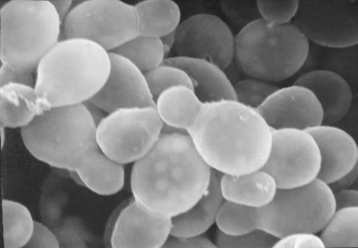
Pityriasis versicolor is a skin condition that is caused by a type of yeast called Malassezia furfur, which has an affinity for lipids. Malassezia furfur is a constituent of the typical microbial community and is spread through direct physical contact. Pityriasis versicolor is a prevalent infection that is found globally, but is more common in tropical regions. It is also more prevalent among individuals who have received a kidney transplant and those with AIDS.
Pityriasis versicolor presents as desiccated, flaky, hyperpigmented, or hypopigmented patches on the trunk, upper limbs, and midsection. Malassezia furfur has the potential to induce systemic illness in individuals undergoing intravenous intralipid treatment, as well as seborrheic dermatitis in patients with AIDS.
Pityriasis versicolor is not linked to a host immune response, and there are no known virulence factors associated with it.
Upon direct microscopic observation of scaly lesions in a KOH preparation, a distinctive "spaghetti and meatballs" look of yeast and hyphae can be observed.
Pityriasis versicolor is commonly treated using topical applications of selenium sulfide or miconazole. Maintaining good cleanliness is crucial for preventing diseases.
Pityriasis versicolor is a skin condition that is caused by a type of yeast called Malassezia furfur, which has an affinity for lipids. Malassezia furfur is a constituent of the typical microbial community and is spread through direct physical contact. Pityriasis versicolor is a prevalent infection that is found globally, but is more common in tropical regions. It is also more prevalent among individuals who have received a kidney transplant and those with AIDS.
Pityriasis versicolor presents as desiccated, flaky, hyperpigmented, or hypopigmented patches on the trunk, upper limbs, and midsection. Malassezia furfur has the potential to induce systemic illness in individuals undergoing intravenous intralipid treatment, as well as seborrheic dermatitis in patients with AIDS.
Pityriasis versicolor is not linked to a host immune response, and there are no known virulence factors associated with it.
Upon direct microscopic observation of scaly lesions in a KOH preparation, a distinctive "spaghetti and meatballs" look of yeast and hyphae can be observed.
Pityriasis versicolor is commonly treated using topical applications of selenium sulfide or miconazole. Maintaining good cleanliness is crucial for preventing diseases.

- Published on
Infectious Diseases and Microbiology - Tinea Nigra ( Exophilia werneckii)
Tinea nigra is a fungal infection caused by Exophilia werneckii, which is a type of fungus that can produce melanin and has two different forms. E werneckii is commonly present in the soil and is frequently transferred through physical harm. Tinea nigra is prevalent in tropical regions.
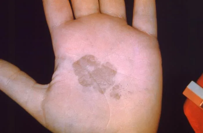
Tinea nigra is distinguished by the presence of smooth, brown to black patches on the palms, soles, and occasionally other areas of the body.
The development and progression of a disease. Tinea nigra does not provoke an immunological response in the host, and there are no recognized mechanisms that contribute to its pathogenicity.
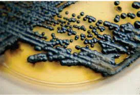
Microscopic analysis of skin scrapings using a KOH preparation indicates the presence of brown colored yeast cells and hyphae. The culture confirmation indicates the presence of black colonies.Topical use of salicylic acid is used to treat tinea nigra. Maintaining good hygiene is a crucial preventive measure.
Tinea nigra is a fungal infection caused by Exophilia werneckii, which is a type of fungus that can produce melanin and has two different forms. E werneckii is commonly present in the soil and is frequently transferred through physical harm. Tinea nigra is prevalent in tropical regions.
Tinea nigra is distinguished by the presence of smooth, brown to black patches on the palms, soles, and occasionally other areas of the body.
The development and progression of a disease. Tinea nigra does not provoke an immunological response in the host, and there are no recognized mechanisms that contribute to its pathogenicity.
Microscopic analysis of skin scrapings using a KOH preparation indicates the presence of brown colored yeast cells and hyphae. The culture confirmation indicates the presence of black colonies.Topical use of salicylic acid is used to treat tinea nigra. Maintaining good hygiene is a crucial preventive measure.
- Published on
Infectious Diseases and Microbiology - White Piedra ( Trichosporon asahii)
White Piedra is a fungal infection that affects the hair shafts, causing the formation of white or light-colored nodules.
White piedra is a fungal infection that affects the scalp, mustache, beard, or pubic hair. It is caused by the Trichosporon asahii fungus (formerly known as T beigelii). Black piedra is a scalp hair infection caused by the fungus Piedraia hortae.
White and black piedra can be transmitted through infected hairs on combs or hairbrushes that are shared. White and black piedra are prevalent among young adults residing in tropical regions.
White piedra is distinguished by the presence of pale-colored, pliable nodules on the hair shaft. Black piedra is distinguished by the presence of firm, brown to black nodules that are firmly linked to the hair shaft. T asahii has the potential to cause disseminated trichosporonosis, particularly in individuals who have low levels of neutrophils and impaired immune systems.
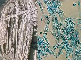
Both white and black piedra do not trigger an immunological response in the host, and there are no recognized variables that contribute to their pathogenicity.Microscopic analysis of hairs in a KOH preparation shows the presence of white to light brown nodules with septate hyphae on the hair shaft, indicating white piedra. Alternatively, dark pigmented nodules containing dark septate hyphae on the hair shaft indicate black piedra.
Piedra is managed with depilation of the affected hairs together with maintaining proper personal cleanliness.
Prophylaxis
White Piedra is a fungal infection that affects the hair shafts, causing the formation of white or light-colored nodules.
White piedra is a fungal infection that affects the scalp, mustache, beard, or pubic hair. It is caused by the Trichosporon asahii fungus (formerly known as T beigelii). Black piedra is a scalp hair infection caused by the fungus Piedraia hortae.
White and black piedra can be transmitted through infected hairs on combs or hairbrushes that are shared. White and black piedra are prevalent among young adults residing in tropical regions.
White piedra is distinguished by the presence of pale-colored, pliable nodules on the hair shaft. Black piedra is distinguished by the presence of firm, brown to black nodules that are firmly linked to the hair shaft. T asahii has the potential to cause disseminated trichosporonosis, particularly in individuals who have low levels of neutrophils and impaired immune systems.
Both white and black piedra do not trigger an immunological response in the host, and there are no recognized variables that contribute to their pathogenicity.Microscopic analysis of hairs in a KOH preparation shows the presence of white to light brown nodules with septate hyphae on the hair shaft, indicating white piedra. Alternatively, dark pigmented nodules containing dark septate hyphae on the hair shaft indicate black piedra.
Piedra is managed with depilation of the affected hairs together with maintaining proper personal cleanliness.
Prophylaxis

- Published on
Infectious Diseases and Microbiology - Tinea Capitis ( Trichosporon tonsurans and Microsporum canis)
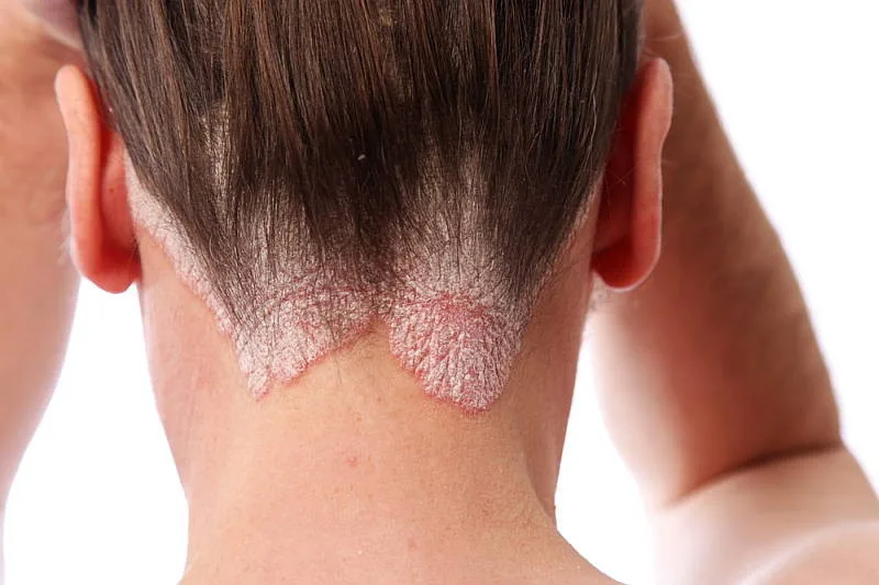
Tinea Capitis, often known as Ringworm, is a fungal infection that affects the scalp.
Tinea capitis, also known as scalp ringworm, is mostly caused by the fungi Trichophyton tonsurans and the zoophilic Microsporum canis. Tinea infections are spread through direct contact with infected individuals or animals (zoophilic species), or indirectly through contact with shed skin or hair on objects including clothing, towels, combs, and brushes. Tinea capitis primarily affects children and has the potential to spread quickly within a family or school setting. Onychomycosis, which is a fungal infection of the nails caused by dermatophytes, impacts approximately 20% of adults in the United States and has a notable negative impact on social well-being.
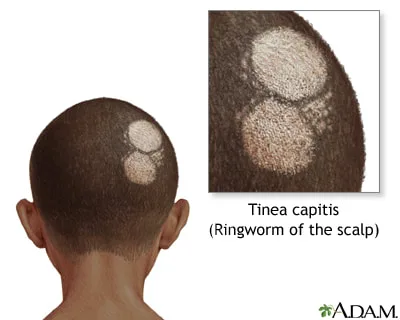
Tinea capitis is a condition that is identified by dry, round, scaly, itchy, red lesions on the scalp. It can also cause patches of hair loss.
Dermatophytes are fungi that have an affinity for keratin and cause infections that are limited to the skin, hair, and nails. Dermatophytes produce keratinase, an enzyme that breaks down keratin and triggers an inflammatory reaction that restricts the spread of infection.
The occurrence of deeper tissue invasion is infrequent due to the nonspecific host defense mechanisms. Individuals with impairments in cell-mediated immunity are susceptible to chronic or widespread dermatophyte infections.
Microscopic analysis of skin, hair, or nails using a KOH preparation reveals the presence of hyphae and diagnostic conidia that are specific to the dermatophyte. Some fungus, specifically Microsporum, exhibit fluorescence when exposed to ultraviolet (UV) light.
Dermatophyte infections are commonly treated using topical applications of tolnaftate and terbinafine. Systemic treatment for onychomycosis involves the administration of either itraconazole or terbinafine. Maintaining proper personal hygiene is crucial for prevention.
Written observations or records of information. Additional types of tinea infections include tinea cruris, commonly known as "jock itch," which is typically caused by Trichophyton rubrum or Epidermophyton floccosum. Tinea pedis, also known as "athlete's foot," is usually caused by T rubrum, T mentagrophytes, or E floccosum. Tinea barbae is typically caused by the zoophilic T verrucosum. Tinea unguium, or onychomycosis, is usually caused by T rubrum.
Tinea Capitis, often known as Ringworm, is a fungal infection that affects the scalp.
Tinea capitis, also known as scalp ringworm, is mostly caused by the fungi Trichophyton tonsurans and the zoophilic Microsporum canis. Tinea infections are spread through direct contact with infected individuals or animals (zoophilic species), or indirectly through contact with shed skin or hair on objects including clothing, towels, combs, and brushes. Tinea capitis primarily affects children and has the potential to spread quickly within a family or school setting. Onychomycosis, which is a fungal infection of the nails caused by dermatophytes, impacts approximately 20% of adults in the United States and has a notable negative impact on social well-being.
Tinea capitis is a condition that is identified by dry, round, scaly, itchy, red lesions on the scalp. It can also cause patches of hair loss.
Dermatophytes are fungi that have an affinity for keratin and cause infections that are limited to the skin, hair, and nails. Dermatophytes produce keratinase, an enzyme that breaks down keratin and triggers an inflammatory reaction that restricts the spread of infection.
The occurrence of deeper tissue invasion is infrequent due to the nonspecific host defense mechanisms. Individuals with impairments in cell-mediated immunity are susceptible to chronic or widespread dermatophyte infections.
Microscopic analysis of skin, hair, or nails using a KOH preparation reveals the presence of hyphae and diagnostic conidia that are specific to the dermatophyte. Some fungus, specifically Microsporum, exhibit fluorescence when exposed to ultraviolet (UV) light.
Dermatophyte infections are commonly treated using topical applications of tolnaftate and terbinafine. Systemic treatment for onychomycosis involves the administration of either itraconazole or terbinafine. Maintaining proper personal hygiene is crucial for prevention.
Written observations or records of information. Additional types of tinea infections include tinea cruris, commonly known as "jock itch," which is typically caused by Trichophyton rubrum or Epidermophyton floccosum. Tinea pedis, also known as "athlete's foot," is usually caused by T rubrum, T mentagrophytes, or E floccosum. Tinea barbae is typically caused by the zoophilic T verrucosum. Tinea unguium, or onychomycosis, is usually caused by T rubrum.
- Published on
Infectious Diseases and Microbiology - Sporotrichosis ( Sporothrix schenckii)
Sporotrichosis is a fungal infection.
Sporotrichosis is a result of infection by the dimorphic fungus Sporothrix schenckii. Sporotrichosis is contracted through occupational or recreational contact with the organism, which is present in the soil or decaying vegetation, typically by means of a puncture wound such as a thorn prick. Gardeners often get sporotrichosis. Rose thorns, sphagnum moss, and baled hay can serve as sources of infection.
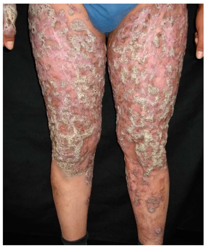
Sporotrichosis presents as nodules and ulcers at the location of infection and along the lymphatic pathway.
Sporotrichosis is a condition characterized by a confined infection that triggers an inflammatory reaction. The synthesis of melanin by S schenckii may impede the destruction caused by neutrophils. Cell-mediated immunity is the primary method by which the host defends itself against pathogens.
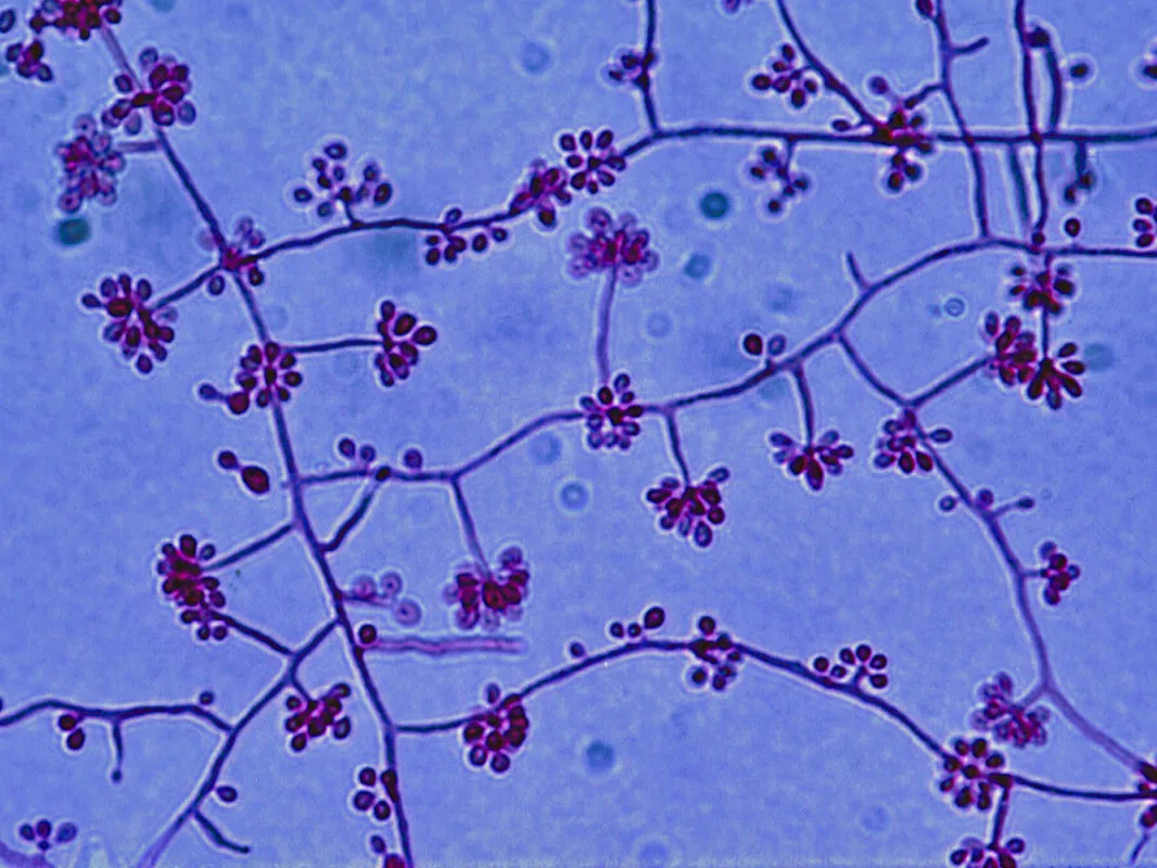
The diagnosis is established through the evidence of dimorphism, where a mold is observed at 25°C and yeast at 37°C, using culture techniques.
The treatment for sporotrichosis involves oral administration of potassium iodide (less frequently used) or systemic administration of itraconazole or amphotericin B. Precautionary precautions involve using gloves and protective attire while dealing with rose bushes, sphagnum moss, hay bales, and wood splinters.
Sporotrichosis is a fungal infection.
Sporotrichosis is a result of infection by the dimorphic fungus Sporothrix schenckii. Sporotrichosis is contracted through occupational or recreational contact with the organism, which is present in the soil or decaying vegetation, typically by means of a puncture wound such as a thorn prick. Gardeners often get sporotrichosis. Rose thorns, sphagnum moss, and baled hay can serve as sources of infection.
Sporotrichosis presents as nodules and ulcers at the location of infection and along the lymphatic pathway.
Sporotrichosis is a condition characterized by a confined infection that triggers an inflammatory reaction. The synthesis of melanin by S schenckii may impede the destruction caused by neutrophils. Cell-mediated immunity is the primary method by which the host defends itself against pathogens.
The diagnosis is established through the evidence of dimorphism, where a mold is observed at 25°C and yeast at 37°C, using culture techniques.
The treatment for sporotrichosis involves oral administration of potassium iodide (less frequently used) or systemic administration of itraconazole or amphotericin B. Precautionary precautions involve using gloves and protective attire while dealing with rose bushes, sphagnum moss, hay bales, and wood splinters.
- Published on
Infectious Diseases and Microbiology - Chromoblastomycosis ( Fonsecaea, Cladosporium, Phialophora)
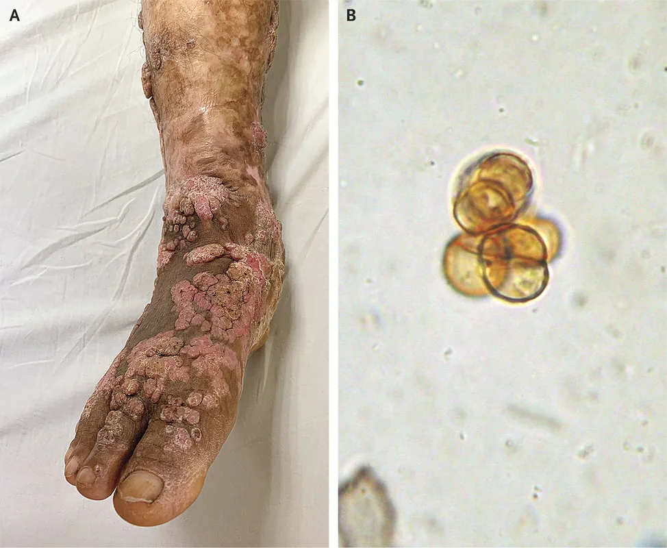
Chromoblastomycosis is a fungal infection caused by many types of soil fungi (Fonsecaea, Cladosporium, Phialophora) that are commonly found in tropical regions. This infection is infrequently observed in the United States. The majority of infections typically manifest in the feet and lower extremities of those who do not wear footwear.
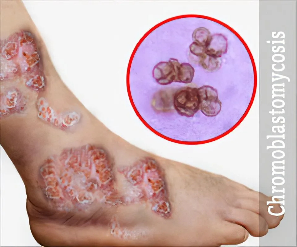
Chromoblastomycosis is defined by the gradual formation of wartlike growths that eventually transform into a cauliflower-like appearance at the site of infection.
Chromoblastomycosis lesions display an inflammatory reaction characterized by the presence of keratinolytic microabscesses and an increase in the number of epithelial cells. The examination of skin scrapings in a KOH preparation shows the presence of copper-colored cells known as sclerotic (muriform) bodies, which are highly indicative of chromoblastomycosis.
Chromoblastomycosis can be treated through surgical removal or with the administration of flucytosine or itraconazole. Wearing shoes is a precautionary measure.
Chromoblastomycosis is a fungal infection caused by many types of soil fungi (Fonsecaea, Cladosporium, Phialophora) that are commonly found in tropical regions. This infection is infrequently observed in the United States. The majority of infections typically manifest in the feet and lower extremities of those who do not wear footwear.
Chromoblastomycosis is defined by the gradual formation of wartlike growths that eventually transform into a cauliflower-like appearance at the site of infection.
Chromoblastomycosis lesions display an inflammatory reaction characterized by the presence of keratinolytic microabscesses and an increase in the number of epithelial cells. The examination of skin scrapings in a KOH preparation shows the presence of copper-colored cells known as sclerotic (muriform) bodies, which are highly indicative of chromoblastomycosis.
Chromoblastomycosis can be treated through surgical removal or with the administration of flucytosine or itraconazole. Wearing shoes is a precautionary measure.