- Published on
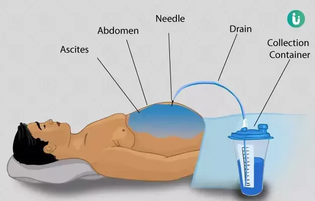
Clinical Procedures - Ascitic Fluid Sampling (Tap)
Indications
• Diagnosing nature of new onset ascites (i.e. exudate or transudate)
• Diagnosis of spontaneous bacterial peritonitis
• Cytology to diagnose malignant :
ascites.
Contraindications
• Acute abdomen that requires surgery
• Pregnancy
• Intestinal obstruction
• Grossly distended urinary bladder
• Superficial infection (cellulitis) at the potential puncture site
• Hernia at the potential puncture site.
Risks
• Persistent leak of ascitic fluid:
• This is more likely if there is a large amount of fluid under tension
• Perforation of hollow viscera (e.g. bowel and bladder). This is very rare
• Peritonitis
• Abdominal wall hematoma
• Bleeding is very rare but may occur if there is injury to inferior epigastric artery (be careful to tap lateral abdominal wall as described).
Procedure Tips
• Check the patient's clotting and platelet count before the procedure and proceed with caution and senior advice if abnormal (correct if platelets <20 x 10°L, INR ≥2.5)
• Inform the laboratory especially during out of hours if cultures needed urgently and if SBP is suspected
• D If unable to obtain fluid despite correct technique, do not persist!
Stop and seek senior advice.
SAAG
Calculating the serum-ascites albumin gradient (SAAG) is a method of determining the cause of ascites.
SAAG = [serum albumin] - [ascites albumin]
>11g/L = portal hypertension (cirrhosis, alcoholic hepatitis, cardiac
ascites, Budd-Chiari syndrome, massive liver metastases).
<11g/L = infection, malignancy, nephrotic syndrome, pancreatic ascites.
Equipment
• Sterile gloves
• Dressing pack
• Antiseptic solution (e.g. iodine)
• 1% or 2% lidocaine
• 1 × 20mL syringe
• 2 × 5mL syringes
• 21G (green) and 25G (orange) needles
• Sterile containers
• Culture bottles
• Sterile dressing.
Procedure
• Introduce yourself, confirm the patient's identity, explain the procedure, and obtain informed consent
• Examine the abdomen and select a site for aspiration, 3 finger-breadths cranial to the anterior superior iliac spine:
• D Beware of positioning too medial as this risks hitting the inferior epigastric vessels
• D Be sure to identify and avoid any organomegaly which might interfere with procedure (in patients with massive splenomegaly, for example, avoid left iliac fossa)
• Clean the area with disinfectant and apply sterile drape
• Using the 25G (orange) needle and the 5mL syringe, administer local anaesthetic to the skin and subcutis, raising a wheal
• Using the 21G (green) needle, infiltrate deeper tissues, intermittently applying suction until the peritoneal cavity is reached, confirmed by flow of ascitic fluid into the syringe
• Note the depth needed to enter the peritoneal cavity
• Discard the used needles and attach a clean 21G needle to the 20mL syringe
• With the green needle perpendicular to the skin, insert carefully, aspirating continuously until you feel resistance give way
• Aspirate as much fluid as needed (usually 20mL is plenty)
• Withdraw needle and syringe and apply dressing
•Send sample for Gram stain and culture (in blood culture bottles), white cell count/neutrophils, biochemistry, cytology (if malignancy suspected):
• White cell count can be calculated in haematology lab; send fluid in EDTA-containing bottle. » (Total white cell count >500/mm' or neutrophils >250/mm3 suggests spontaneous bacterial peritonitis, SP)
• Neutrophil count is usually a manual procedure via microbiology and may take longer
• If malignancy is suspected, a large volume of ascites (e.g.
500mL) should be sent to cytology.
Documentation
• Date, time, indication, informed consent obtained
• Type and amount of local anaesthetic used
• Site aspirated
• Aseptic technique used?
• How many passes?
• Volume and colour of aspirate obtained
• Tests requested on samples
• Any immediate complications
• Signature, printed name, and contact details.
Indications
• Diagnosing nature of new onset ascites (i.e. exudate or transudate)
• Diagnosis of spontaneous bacterial peritonitis
• Cytology to diagnose malignant :
ascites.
Contraindications
• Acute abdomen that requires surgery
• Pregnancy
• Intestinal obstruction
• Grossly distended urinary bladder
• Superficial infection (cellulitis) at the potential puncture site
• Hernia at the potential puncture site.
Risks
• Persistent leak of ascitic fluid:
• This is more likely if there is a large amount of fluid under tension
• Perforation of hollow viscera (e.g. bowel and bladder). This is very rare
• Peritonitis
• Abdominal wall hematoma
• Bleeding is very rare but may occur if there is injury to inferior epigastric artery (be careful to tap lateral abdominal wall as described).
Procedure Tips
• Check the patient's clotting and platelet count before the procedure and proceed with caution and senior advice if abnormal (correct if platelets <20 x 10°L, INR ≥2.5)
• Inform the laboratory especially during out of hours if cultures needed urgently and if SBP is suspected
• D If unable to obtain fluid despite correct technique, do not persist!
Stop and seek senior advice.
SAAG
Calculating the serum-ascites albumin gradient (SAAG) is a method of determining the cause of ascites.
SAAG = [serum albumin] - [ascites albumin]
>11g/L = portal hypertension (cirrhosis, alcoholic hepatitis, cardiac
ascites, Budd-Chiari syndrome, massive liver metastases).
<11g/L = infection, malignancy, nephrotic syndrome, pancreatic ascites.
Equipment
• Sterile gloves
• Dressing pack
• Antiseptic solution (e.g. iodine)
• 1% or 2% lidocaine
• 1 × 20mL syringe
• 2 × 5mL syringes
• 21G (green) and 25G (orange) needles
• Sterile containers
• Culture bottles
• Sterile dressing.
Procedure
• Introduce yourself, confirm the patient's identity, explain the procedure, and obtain informed consent
• Examine the abdomen and select a site for aspiration, 3 finger-breadths cranial to the anterior superior iliac spine:
• D Beware of positioning too medial as this risks hitting the inferior epigastric vessels
• D Be sure to identify and avoid any organomegaly which might interfere with procedure (in patients with massive splenomegaly, for example, avoid left iliac fossa)
• Clean the area with disinfectant and apply sterile drape
• Using the 25G (orange) needle and the 5mL syringe, administer local anaesthetic to the skin and subcutis, raising a wheal
• Using the 21G (green) needle, infiltrate deeper tissues, intermittently applying suction until the peritoneal cavity is reached, confirmed by flow of ascitic fluid into the syringe
• Note the depth needed to enter the peritoneal cavity
• Discard the used needles and attach a clean 21G needle to the 20mL syringe
• With the green needle perpendicular to the skin, insert carefully, aspirating continuously until you feel resistance give way
• Aspirate as much fluid as needed (usually 20mL is plenty)
• Withdraw needle and syringe and apply dressing
•Send sample for Gram stain and culture (in blood culture bottles), white cell count/neutrophils, biochemistry, cytology (if malignancy suspected):
• White cell count can be calculated in haematology lab; send fluid in EDTA-containing bottle. » (Total white cell count >500/mm' or neutrophils >250/mm3 suggests spontaneous bacterial peritonitis, SP)
• Neutrophil count is usually a manual procedure via microbiology and may take longer
• If malignancy is suspected, a large volume of ascites (e.g.
500mL) should be sent to cytology.
Documentation
• Date, time, indication, informed consent obtained
• Type and amount of local anaesthetic used
• Site aspirated
• Aseptic technique used?
• How many passes?
• Volume and colour of aspirate obtained
• Tests requested on samples
• Any immediate complications
• Signature, printed name, and contact details.
0 Comments
