- Published on
Clinical Procedures - Non Invasive Ventilation ( CPAP)
Non-invasive ventilation should only be set up by experienced operators. The following is a guide only.
Background
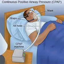
CAP = continuous positive airways pressure. CAP traditionally
has its own equipment and 'set-up'. Recently more clinicians are delivering CPAP through the BiPAP Vision@. There is also a
'low-flow' version used mainly for transport of CPAP-dependent patients.
Equipment
)rd
• Mask (‡ T-piece), hood
• Head strap (mask), shoulder straps (hood)
• Oxygen circuit and humidification
• High flow generator (e.g. Whisper Flow®, Vital Signs®)
• Positive end-expiratory pressure (PEEP) valves (usually 5, 7.5, or 10cmH,O)
• 'Blow off' safety valve (10cmHO above the PEEP used).
Procedure
• Use available templates to assess appropriate sized interface and minimize air leaks (if using the BiPAP Vision
• Decide on level of PEEP to apply
• Attach PEEP valve to mask (if using traditional set-up, may need T-piece)
• Attach oxygen circuit with humidification including 'blow off' valve (for safety)
• Set inspired oxygen level
• Set flow rate to ensure the PEEP valve opens a small distance and never closes
• Titrate oxygen and PEEP in response to the patient's work of breathing, saturations, pH, Pa02, and PaCO2
• If appropriate, set alarms on ventilator (if using BiPAP Vision®
• Write a prescription chart of PEEP or ventilation settings and acceptable saturations, Pa02 and PaCO, continuous or intermittent.
Procedure Tips
• If valve closing fully, there is an air leak or patient requires higher PEEP
• May need to put more air into the mask rim for patient comfort and a better seal
• Better fit of mask if the patient keeps their dentures in!
• Large-bore nasogastric tubes will cause a leak, change to small-bore or bulk up cheek tissue with granuflex.
Indications
• Type 1 respiratory failure (e.g. lobar or bibasal collapse post surgery, pneumonia)
• Cardiogenic pulmonary edema
• Obstructive sleep apnoea
• Weaning.
Contraindications /Cautions
• D Undrained pneumothorax (absolute contraindication)
• Facial fractures
• Life-threatening epistaxis
• Bullous pulmonary disease
• Proximal lung tumours (air trapping)
• Active TB (spread)
• Acute head injury
• Low blood pressure
• Uncontrolled cardiac arrhythmias
• Sinus/middle ear infection.
Risks
• Abdominal distension (secondary to 'swallowing' air)
• Decreased cardiac output (drop in blood pressure)
• Pressure sores from mask
• Aspiration of vomit
• CO, retention if patient breathing small tidal volume against
Documentation
• Oxygen prescription chart
• Clear record of ABGs with evidence of time, inspired oxygen and PEEP
• Continuous or intermittent
• Good practice to document 'ceiling' of treatment in clinical environment (pressure and Fi02).
Non-invasive ventilation should only be set up by experienced operators. The following is a guide only.
Background
CAP = continuous positive airways pressure. CAP traditionally
has its own equipment and 'set-up'. Recently more clinicians are delivering CPAP through the BiPAP Vision@. There is also a
'low-flow' version used mainly for transport of CPAP-dependent patients.
Equipment
)rd
• Mask (‡ T-piece), hood
• Head strap (mask), shoulder straps (hood)
• Oxygen circuit and humidification
• High flow generator (e.g. Whisper Flow®, Vital Signs®)
• Positive end-expiratory pressure (PEEP) valves (usually 5, 7.5, or 10cmH,O)
• 'Blow off' safety valve (10cmHO above the PEEP used).
Procedure
• Use available templates to assess appropriate sized interface and minimize air leaks (if using the BiPAP Vision
• Decide on level of PEEP to apply
• Attach PEEP valve to mask (if using traditional set-up, may need T-piece)
• Attach oxygen circuit with humidification including 'blow off' valve (for safety)
• Set inspired oxygen level
• Set flow rate to ensure the PEEP valve opens a small distance and never closes
• Titrate oxygen and PEEP in response to the patient's work of breathing, saturations, pH, Pa02, and PaCO2
• If appropriate, set alarms on ventilator (if using BiPAP Vision®
• Write a prescription chart of PEEP or ventilation settings and acceptable saturations, Pa02 and PaCO, continuous or intermittent.
Procedure Tips
• If valve closing fully, there is an air leak or patient requires higher PEEP
• May need to put more air into the mask rim for patient comfort and a better seal
• Better fit of mask if the patient keeps their dentures in!
• Large-bore nasogastric tubes will cause a leak, change to small-bore or bulk up cheek tissue with granuflex.
Indications
• Type 1 respiratory failure (e.g. lobar or bibasal collapse post surgery, pneumonia)
• Cardiogenic pulmonary edema
• Obstructive sleep apnoea
• Weaning.
Contraindications /Cautions
• D Undrained pneumothorax (absolute contraindication)
• Facial fractures
• Life-threatening epistaxis
• Bullous pulmonary disease
• Proximal lung tumours (air trapping)
• Active TB (spread)
• Acute head injury
• Low blood pressure
• Uncontrolled cardiac arrhythmias
• Sinus/middle ear infection.
Risks
• Abdominal distension (secondary to 'swallowing' air)
• Decreased cardiac output (drop in blood pressure)
• Pressure sores from mask
• Aspiration of vomit
• CO, retention if patient breathing small tidal volume against
Documentation
• Oxygen prescription chart
• Clear record of ABGs with evidence of time, inspired oxygen and PEEP
• Continuous or intermittent
• Good practice to document 'ceiling' of treatment in clinical environment (pressure and Fi02).
0 Comments
