- Published on
Kembara Xtra - Medicine - Pressure Ulcer
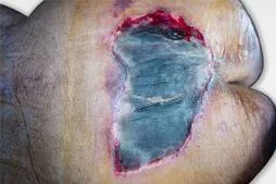
A specific area of skin or underlying tissue damage brought on by pressure or shear
.Typically over a bony prominence (such as the sacrum, calcaneus, or ischium), and is categorized by the National Pressure Injury Advisory Panel (NPIAP) into the following stages:
- Stage I: nonblanchable erythema, in which the skin is still present but has a nonblanchable redness; darkly pigmented skin may not exhibit obvious blanching.
Stage II: Partial-thickness skin loss; intact or open/ruptured serum-filled blisters; shallow open ulcer with a viable reddish-pink, wet wound bed; without slough.
- Stage III: complete loss of skin thickness; subcutaneous fat may be apparent, but bone, tendon, or muscle are not exposed; the presence of slough, if any, does not conceal the extent of tissue loss.
Stage IV: full-thickness tissue loss, including exposed bone, tendon, or joint; slough or eschar may be present but does not entirely cover the wound base.
- Unstageable: depth is uncertain; slough and/or eschar in the wound bed conceal the ulcer's base.
- Suspected deep tissue injury: unclear depth, maroon or purple region of intact skin, or blister filled with blood. Skin color changes are preceded by pain and temperature changes.
Synonyms include bedsore, pressure sore, and decubitus ulcer.
Prevalence depends on setting and population and is dependent on epidemiology incidence (0–53.4%–0–72.5%).
Pathophysiology and Etiology
Complex interaction between risk variables and outside forces (such as pressure and/or shear, friction, and moisture)
Risk factors include reduced skin perfusion, malnutrition, impaired mobility, sensory impairment, and the use of medical devices.
Repositioning, early mobilization, support surfaces, microclimate control, prophylactic dressings, structured risk assessment, skin and tissue assessment, preventive skin care, nutrition screening, and electrical stimulation of the muscles are all examples of general prevention.
Elderly age, immobility, trauma, hip fractures, diabetes, cerebrovascular and cardiovascular disease, and incontinence are all associated conditions.
Information about the diagnosis, risk factors, nutritional assessment, pain assessment, date of ulcer diagnosis, and course of treatment.
clinical assessment
A thorough skin inspection upon first meeting and at intervals throughout visits
Determine the presence of sinus tracts, undermining, tunneling, exudate, necrosis, odor, and evidence of healing (such as granulation tissue), and evaluate the location, stage, size (length, width, and depth).
Determine the elements (impaired perfusion, sensibility, infection) that may have an impact on the healing process.
Cellulitis and erysipelas, incontinence-related dermatitis, skin tears, intertrigo, neuropathic ulcers, cutaneous squamous cell carcinomas, hypertensive ulcers, pyoderma gangrenosum, cancers, vasculitides, and other dermatologic conditions are among the differential diagnoses.
Laboratory Results
Initial examinations (lab, imaging)
Culture of wounds: Avoid culture of surface drainage. Perform a bone biopsy and deep tissue culture if necessary.
Add infectious workup, including inflammatory markers, CBC, blood cultures, and x-rays if systemic infection or that of bone or muscle is suspected. To confirm osteomyelitis, an MRI might be required.
Doppler ultrasound and the ankle-brachial index (for wounds on the lower extremities)
Tests in the Future & Special Considerations
When a different medical condition makes assessment more difficult, extra testing could be recommended. This could involve performing tests for dermatological illnesses such as diabetes (i.e., A1c), thyroid disease (i.e., TSH), vascular disease, and others.
Management
A thorough initial evaluation of the patient; a lowering or redistribution of pressure; and nutritional support (such as protein-containing supplements)
The preparation of the wound bed (tissue management, infection and inflammation control, moisture balancing, advancement of the epithelial edge) is part of the wound assessment and treatment process.- Debridement - Cleaning of the wound
Address stiffness.
Manage incontinence, deal with pain, and evaluate care objectives and advance directives.
First Line of Medicine
Dressings for wounds that are suitable for the kind of ulcer (transparent film, hydrocolloid, hydrogel, alginate, foam, silver-impregnated, honey-impregnated, cadexomer iodine, gauze, silicone, collagen matrix, composite).
Systematic reviews uncover evidence that one dressing is preferable to another that is of subpar quality.
As a result, recommending one dressing over another is impossible.
Agents for enzymatic débridement
Next Line
Charcoal, activated
Topical antiseptics, such as hydrogen peroxide, the Dakin solution, and povidone-iodine
Referral
Consider referring patients with complex or slow-healing wounds to a wound care specialist (if one is available).
If necessary, take into account vascular surgery to enhance the blood flow to the wound via a vascular bypass.
Consider seeking a surgical opinion if cellulitis is progressing, if sepsis is suspected to be the cause, if there is extensive necrotic tissue that cannot be removed using nonsurgical débridement techniques, or if stage III or IV lesions are not improving with conservative treatment.
If necessary, think about plastic surgery for a skin graft or flap.
Consider referring someone to a dermatologist if pyoderma gangrenosum, malignancy, vasculitis, or other dermatological diseases are suspected.
Further Treatments
Direct contact electrical stimulation for resistant stage II, any category, stage III, and stage IV Electromagnetic field for any stage III or stage IV stubborn subject in category/stage II Pulsed radio frequency radiation for any stage III or IV that is recalcitrant, as well as stage II.
Alternative Therapies
• High-frequency ultrasound as an adjuvant for infected pressure ulcers • Low-frequency ultrasound for débridement of necrotic soft tissue (not eschar) Early adjuvant therapy using negative pressure wound care for deep, stage III, and stage IV wounds
Consider a course of hydrotherapy with pulsed lavage and suction for wound cleaning and débridement. If conventional therapies are unsuccessful, try phototherapy with short-term UVC light. Laser and infrared are not advised.
Consider maggot débridement therapy instead of hyperbaric and topical oxygen therapy, vibration therapy, hydrotherapy with a whirlpool, and hyperbaric and topical oxygen therapy.
Admission
Dressing changes 1 to 3 times per day based on wound assessment and care plan Admission criteria/initial stabilization: refractory cellulitis, osteomyelitis, systemic infection, advanced nutritional deterioration, suspected patient abuse, incapacity to care for oneself
Assessing risk factors in accordance with scales; checking for new or changing wounds; and determining the safest and most appropriate place to discharge patients are the criteria for discharge.
Follow-Up Evaluation by a nurse with wound experience; biweekly evaluation by a doctor
Patient monitoring, home health nursing, and a change in the plan of care if there is no improvement after two to three weeks.
Diet: Approximately 1.0 to 1.5 kg/day of protein; strict glycemic management; addition of micronutrients (such as vitamin C and zinc) to the diet or as supplements.
Patient Education Regularly examine your skin.
Infection symptoms Report any new or worsening pain.
Prevention of new wounds where previous wounds have healed, prevention of dampness, and quitting smoking
The following factors will affect the prognosis: pressure relief, nutrition, and wound care.
Increased mortality, infections, complications, and amputations

0 Comments
