- Published on
Pathology - Idiopathic Pulmonary Fibrosis
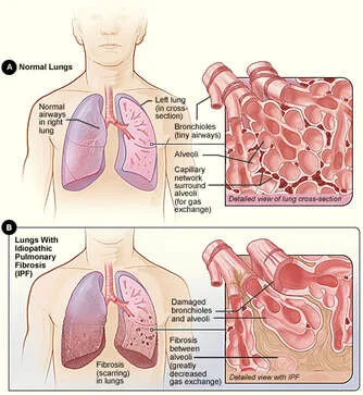
Idiopathic Pulmonary Fibrosis is a condition characterized by fibrosis of the lungs, and its cause is still unknown. A proposed process involves the initial insult to the alveolar epithelium, which subsequently triggers abnormal activation of fibroblasts, resulting in damage to the lung tissue.
The microscopic examination reveals a significant increase in fibrous tissue and the growth of fibroblast cells, leading to the distortion of the walls separating the air sacs in the lungs. This results in a honeycomb-like structure, with enlarged bronchioles that are lined by scarred and fibrotic lung tissue.
Patients have a gradual onset of difficulty breathing and a non-productive cough.
The physical examination reveals the presence of hypoxemia, inspiratory crackles throughout the lungs, and clubbing.
Imaging: CT scan reveals a subpleural honeycombing pattern and patchy bibasilar reticular opacities.
Pulmonary function test results: Reduced total lung capacity (TLC) and reduced diffusing capacity of the lungs for carbon monoxide (DLCO).
Treatment options include providing additional oxygen and considering the possibility of a lung transplant.
The prognosis is unfavorable, with an average survival rate of 2-5 years following diagnosis.
Idiopathic Pulmonary Fibrosis is a condition characterized by fibrosis of the lungs, and its cause is still unknown. A proposed process involves the initial insult to the alveolar epithelium, which subsequently triggers abnormal activation of fibroblasts, resulting in damage to the lung tissue.
The microscopic examination reveals a significant increase in fibrous tissue and the growth of fibroblast cells, leading to the distortion of the walls separating the air sacs in the lungs. This results in a honeycomb-like structure, with enlarged bronchioles that are lined by scarred and fibrotic lung tissue.
Patients have a gradual onset of difficulty breathing and a non-productive cough.
The physical examination reveals the presence of hypoxemia, inspiratory crackles throughout the lungs, and clubbing.
Imaging: CT scan reveals a subpleural honeycombing pattern and patchy bibasilar reticular opacities.
Pulmonary function test results: Reduced total lung capacity (TLC) and reduced diffusing capacity of the lungs for carbon monoxide (DLCO).
Treatment options include providing additional oxygen and considering the possibility of a lung transplant.
The prognosis is unfavorable, with an average survival rate of 2-5 years following diagnosis.
0 Comments
