- Published on
Pathology - Multiple Myeloma
A tumor cell originates from the rapid growth of a single type of plasma cells, known as monoclonal plasma cells, that are responsible for producing immunoglobulin G (IgG).
Most prevalent among individuals over the age of 60.
Pathology
Bone marrow: Malignant plasma cells with a characteristic fried-egg look originating from B lymphocytes.
Peripheral blood smear reveals the presence of erythrocytes arranged in a structure known as rouleaux.
Long bones: Lytic lesions are caused by the secretion of osteoclast-activating factor by cancerous cells.
Clinical Presentations
Primary amyloidosis is characterized by bone discomfort, fractures, renal failure, and recurrent infections.
Imaging: Visible lytic lesions on radiography
Laboratory results: The patient exhibits a monoclonal immunoglobulin spike (M protein) on serum protein electrophoresis, Bence-Jones protein (IgG light chains) in urine, anemia, elevated erythrocyte sedimentation rate (ESR), and hypercalcemia. Increased levels of immunoglobulin G (IgG) in the blood, together with elevated levels of nitrogenous waste products (azotemia).
Treatment options for patients include chemotherapy, autologous stem cell transplant, and bisphosphonates for those with hypercalcemia.
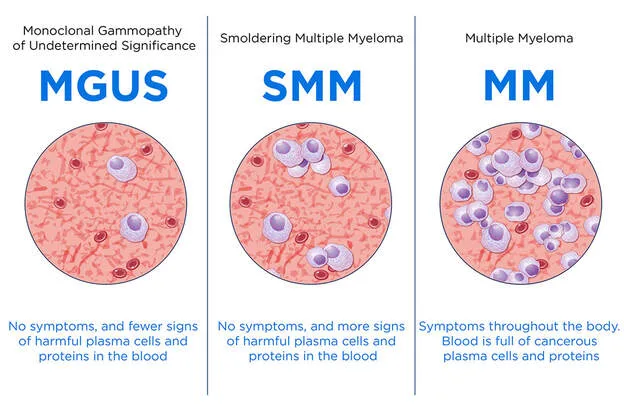
MGUS is a condition without symptoms that is defined by the presence of a monoclonal M protein spike that is less than 3 g/dL. It is not accompanied by Bence-Jones proteinuria, lytic bone lesions, or renal insufficiency. Close monitoring of patients with MGUS is important due to their heightened susceptibility to developing multiple myeloma or another lymphoproliferative illness.
A tumor cell originates from the rapid growth of a single type of plasma cells, known as monoclonal plasma cells, that are responsible for producing immunoglobulin G (IgG).
Most prevalent among individuals over the age of 60.
Pathology
Bone marrow: Malignant plasma cells with a characteristic fried-egg look originating from B lymphocytes.
Peripheral blood smear reveals the presence of erythrocytes arranged in a structure known as rouleaux.
Long bones: Lytic lesions are caused by the secretion of osteoclast-activating factor by cancerous cells.
Clinical Presentations
Primary amyloidosis is characterized by bone discomfort, fractures, renal failure, and recurrent infections.
Imaging: Visible lytic lesions on radiography
Laboratory results: The patient exhibits a monoclonal immunoglobulin spike (M protein) on serum protein electrophoresis, Bence-Jones protein (IgG light chains) in urine, anemia, elevated erythrocyte sedimentation rate (ESR), and hypercalcemia. Increased levels of immunoglobulin G (IgG) in the blood, together with elevated levels of nitrogenous waste products (azotemia).
Treatment options for patients include chemotherapy, autologous stem cell transplant, and bisphosphonates for those with hypercalcemia.
MGUS is a condition without symptoms that is defined by the presence of a monoclonal M protein spike that is less than 3 g/dL. It is not accompanied by Bence-Jones proteinuria, lytic bone lesions, or renal insufficiency. Close monitoring of patients with MGUS is important due to their heightened susceptibility to developing multiple myeloma or another lymphoproliferative illness.
0 Comments
