- Published on
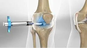
Surgery - Knee Joint Injection
Indications
• Iniection of corticosteroids are performed to relieve:
•Acute attack of crystal arthritis (gout, pseudogout)
• Flare of inflammatory arthritis (seronegative arthritis, rheumatoid arthritis, reactive arthritis)
• Pain in patients with osteoarthritis.
Other substances administered by an intra-articular injection
• Yttrium: for chronic monoarticular synovitis
• Hyaluronic acid: for pain in patients with osteoarthritis of the knee.
Contraindications
• Superficial infection or broken skin
• Coagulopathy: patients with thrombocytopenia and a high IN should be discussed with a rheumatologist or orthopedic surgeon before a joint injection is attempted
• Prosthetic joint.
Risks
• Discomfort: usually short lived and minor
• Septic arthritis: risk is very small (1 in 78,000 in a recent retrospective study from France)
• Subcutaneous atrophy: rare but may be permanent
• Side effects from systemic absorption of corticosteroids: including facial flushing and transient worsening of diabetic control (uncommon)
• Increased pain: this usually lasts for 2-3 days.
Procedure Tips
• It is good practice to send the joint aspirate for routine crystal study Ch stain, and culture. This establishes if the joint was infected before joins aspiration and corticosteroid injection
• Microbiology request forms should provide information on the date, time, and site of joint aspirated.
Injecting smaller joints
• Hydrocortisone (short acting) should be used for small superficial joints rather than triamcinolone
• For MCP joints, 25mg of hydrocortisone and 0.5mL of 2% lidocaine will suffice
• Use 23-25G (blue, orange) for small joints such as wrist, MCP.
Equipment
• Sterile gloves
• Isopropyl alcohol swabs
• Corticosteroid injection:
• Triamcinolone (long acting) for large joints (knee, shoulder)
• Lidocaine 1% or 2%
• 21G (green) needles
• 10mL syringe
• 5mL syringe
• White-capped universal containers
• Elastoplast or cotton wool and sticky tape.
Procedure (Medial Approach)
• Introduce yourself, confirm the patient's identity, explain the procedure, and obtain informed consent
• Position the patient resting on a couch with the leg slightly flexed and supported on a pillow (relaxes quadriceps)
• Identify and mark the point of entry just below the midpoint of the patella by indenting the skin with the tip of a syringe
• Wash hands and put on the gloves
• Draw up 40mg triamcinolone and 2mL of 2% lidocaine
• A no-touch technique is essential after cleaning so any mark to identify the point of entry should be made earlier
• Wipe the site with alcohol swabs
• Attach a 10mL syringe to a green needle
• Insert the needle just below the midpoint of patella, aiming behind the patella. Your free hand can apply pressure to move any synovial fluid to the medial side
• Intra-articular placement of the needle is confirmed by effortless aspiration of synovial fluid
• If the syringe fills up, it can be detached from the needle, the synovial fluid discarded in a specimen pot, and the syringe re-attached to the needle to aspirate more synovial fluid
• If there is no suspicion of septic arthritis and, once a significant proportion of synovial fluid has been aspirated, attach the syringe containing a mixture of corticosteroids and lidocaine to the needle
• Aspirate a little more to confirm intra-articular, extra-vascular placement and inject the steroid mixture
• • If there is any suspicion of septic arthritis, injection of corticosteroids should not be carried out in the same sitting as synovial fluid aspiration:
•Signs and symptoms of septic arthritis may be indolent in the immunosuppressed and a high index of suspicion needs to be maintained. The clinical suspicion of septic arthritis is further strengthened by aspirating cloudy or purulent fluid
• Withdraw the needle and dress the site with a suitable plaster
• Advise the patient to rest the injected area for 24 48 hours.
Procedure (Superolateral Approach)
For large effusions that distend the suprapatellar pouch.
• The needle is introduced above and lateral to the patella at the maximum convexity of the distended pouch
• The needle should be aimed inferiorly and medially.
Documentation
• Date, time, indication, informed consent obtained
• Site and approach taken
• Aseptic technique used?
• How many passes?
• Volume and nature of fluid aspirated and tests requested
• Type and amount of anaesthetic and steroid used
• Any immediate complications
• Signature, printed name, and contact details.
Indications
• Iniection of corticosteroids are performed to relieve:
•Acute attack of crystal arthritis (gout, pseudogout)
• Flare of inflammatory arthritis (seronegative arthritis, rheumatoid arthritis, reactive arthritis)
• Pain in patients with osteoarthritis.
Other substances administered by an intra-articular injection
• Yttrium: for chronic monoarticular synovitis
• Hyaluronic acid: for pain in patients with osteoarthritis of the knee.
Contraindications
• Superficial infection or broken skin
• Coagulopathy: patients with thrombocytopenia and a high IN should be discussed with a rheumatologist or orthopedic surgeon before a joint injection is attempted
• Prosthetic joint.
Risks
• Discomfort: usually short lived and minor
• Septic arthritis: risk is very small (1 in 78,000 in a recent retrospective study from France)
• Subcutaneous atrophy: rare but may be permanent
• Side effects from systemic absorption of corticosteroids: including facial flushing and transient worsening of diabetic control (uncommon)
• Increased pain: this usually lasts for 2-3 days.
Procedure Tips
• It is good practice to send the joint aspirate for routine crystal study Ch stain, and culture. This establishes if the joint was infected before joins aspiration and corticosteroid injection
• Microbiology request forms should provide information on the date, time, and site of joint aspirated.
Injecting smaller joints
• Hydrocortisone (short acting) should be used for small superficial joints rather than triamcinolone
• For MCP joints, 25mg of hydrocortisone and 0.5mL of 2% lidocaine will suffice
• Use 23-25G (blue, orange) for small joints such as wrist, MCP.
Equipment
• Sterile gloves
• Isopropyl alcohol swabs
• Corticosteroid injection:
• Triamcinolone (long acting) for large joints (knee, shoulder)
• Lidocaine 1% or 2%
• 21G (green) needles
• 10mL syringe
• 5mL syringe
• White-capped universal containers
• Elastoplast or cotton wool and sticky tape.
Procedure (Medial Approach)
• Introduce yourself, confirm the patient's identity, explain the procedure, and obtain informed consent
• Position the patient resting on a couch with the leg slightly flexed and supported on a pillow (relaxes quadriceps)
• Identify and mark the point of entry just below the midpoint of the patella by indenting the skin with the tip of a syringe
• Wash hands and put on the gloves
• Draw up 40mg triamcinolone and 2mL of 2% lidocaine
• A no-touch technique is essential after cleaning so any mark to identify the point of entry should be made earlier
• Wipe the site with alcohol swabs
• Attach a 10mL syringe to a green needle
• Insert the needle just below the midpoint of patella, aiming behind the patella. Your free hand can apply pressure to move any synovial fluid to the medial side
• Intra-articular placement of the needle is confirmed by effortless aspiration of synovial fluid
• If the syringe fills up, it can be detached from the needle, the synovial fluid discarded in a specimen pot, and the syringe re-attached to the needle to aspirate more synovial fluid
• If there is no suspicion of septic arthritis and, once a significant proportion of synovial fluid has been aspirated, attach the syringe containing a mixture of corticosteroids and lidocaine to the needle
• Aspirate a little more to confirm intra-articular, extra-vascular placement and inject the steroid mixture
• • If there is any suspicion of septic arthritis, injection of corticosteroids should not be carried out in the same sitting as synovial fluid aspiration:
•Signs and symptoms of septic arthritis may be indolent in the immunosuppressed and a high index of suspicion needs to be maintained. The clinical suspicion of septic arthritis is further strengthened by aspirating cloudy or purulent fluid
• Withdraw the needle and dress the site with a suitable plaster
• Advise the patient to rest the injected area for 24 48 hours.
Procedure (Superolateral Approach)
For large effusions that distend the suprapatellar pouch.
• The needle is introduced above and lateral to the patella at the maximum convexity of the distended pouch
• The needle should be aimed inferiorly and medially.
Documentation
• Date, time, indication, informed consent obtained
• Site and approach taken
• Aseptic technique used?
• How many passes?
• Volume and nature of fluid aspirated and tests requested
• Type and amount of anaesthetic and steroid used
• Any immediate complications
• Signature, printed name, and contact details.
0 Comments
