- Published on
Symptoms and Signs - Differential Diagnosis of Aphthous Stomatitis (Canker Sores)
• White sponge nevus: thick, white corrugated folds involving the buccal mucosa; appears in childhood as an autosomal dominant trait; benign condition
• Varices
• Nicotine stomatitis: whitened palate with red papules
• Melanotic lesions: freckles, lentigines, lentigo, melanoma, Peutz- Jeghers syndrome, Addison’s disease
• Lichen planus: linear, reticular, slightly raised striae on buccal mucosa; skin is involved by pruritic violaceous papules on forearms and inner thighs
• Leukoplakia: white lesions that cannot be scraped off; 20% are premalignant epithelial dysplasia or squamous cell carcinoma
• Leukoedema: filmy opalescent-appearing mucosa, which can be reverted to normal appearance by stretching. This condition is benign.
• Kaposi’s sarcoma: red or purple macules that enlarge to form tumors; seen in patients with AIDS
• Histoplasmosis: ill-defined irregular patch with a granulomatous surface, sometimes ulcerated
• Hemangiomas
• Hairy leukoplakia: shaggy white surface that cannot be wiped off; seen in HIV infection, caused by Epstein-Barr virus
• Erythroplakia: red patch usually caused by epithelial dysplasia or squamous cell carcinoma
• Discoid lupus erythematosus: lesion resembles lichen planus
• Darier’s disease (keratosis follicularis): white papules on the gingivae, alveolar mucosa, and dorsal tongue; skin lesions also present (erythematous papules); inherited as an autosomal
• Coated tongue: accumulation of keratin; harmless condition that can be treated by scraping
• Chemical injury: white sloughing mucosa
• Candidiasis may present with red instead of the more frequent white lesion (see White Lesions). Median rhomboid glossitis is a chronic variant.
• Candidiasis
• Burning tongue (glossopyrosis): normal examination; sometimes associated with denture trauma, anemia, diabetes, vitamin B12 deficiency, psychogenic problems
• Benign migratory glossitis (geographic tongue): area of atrophic depapillated mucosa surrounded by a keratotic border
• Anemia: atrophic reddened glossal mucosa seen with pernicious anemia
• Allergy
• Papilloma
• Verruca vulgaris
• Condyloma acuminatum
• Fibroma
• Epulis
• Pyogenic granuloma
• Mucocele
• Retention cyst
• Primary herpetic gingivostomatitis
• Pemphigus and pemphigoid
• Hand-foot-mouth disease: caused by coxsackievirus group A
• Erythema multiforme
• Herpangina: caused by echovirus
• Traumatic ulcer
• Primary syphilis
• Perlèche (angular cheilitis)
• Recurrent aphthous stomatitis (canker sores)
• Behçet’s syndrome (aphthous ulcers, uveitis, genital ulcerations,
arthritis, and aseptic meningitis)
• Reiter’s syndrome (conjunctivitis, urethritis, and arthritis with
occasional oral ulcerations)
• White sponge nevus: thick, white corrugated folds involving the buccal mucosa; appears in childhood as an autosomal dominant trait; benign condition
• Varices
• Nicotine stomatitis: whitened palate with red papules
• Melanotic lesions: freckles, lentigines, lentigo, melanoma, Peutz- Jeghers syndrome, Addison’s disease
• Lichen planus: linear, reticular, slightly raised striae on buccal mucosa; skin is involved by pruritic violaceous papules on forearms and inner thighs
• Leukoplakia: white lesions that cannot be scraped off; 20% are premalignant epithelial dysplasia or squamous cell carcinoma
• Leukoedema: filmy opalescent-appearing mucosa, which can be reverted to normal appearance by stretching. This condition is benign.
• Kaposi’s sarcoma: red or purple macules that enlarge to form tumors; seen in patients with AIDS
• Histoplasmosis: ill-defined irregular patch with a granulomatous surface, sometimes ulcerated
• Hemangiomas
• Hairy leukoplakia: shaggy white surface that cannot be wiped off; seen in HIV infection, caused by Epstein-Barr virus
• Erythroplakia: red patch usually caused by epithelial dysplasia or squamous cell carcinoma
• Discoid lupus erythematosus: lesion resembles lichen planus
• Darier’s disease (keratosis follicularis): white papules on the gingivae, alveolar mucosa, and dorsal tongue; skin lesions also present (erythematous papules); inherited as an autosomal
• Coated tongue: accumulation of keratin; harmless condition that can be treated by scraping
• Chemical injury: white sloughing mucosa
• Candidiasis may present with red instead of the more frequent white lesion (see White Lesions). Median rhomboid glossitis is a chronic variant.
• Candidiasis
• Burning tongue (glossopyrosis): normal examination; sometimes associated with denture trauma, anemia, diabetes, vitamin B12 deficiency, psychogenic problems
• Benign migratory glossitis (geographic tongue): area of atrophic depapillated mucosa surrounded by a keratotic border
• Anemia: atrophic reddened glossal mucosa seen with pernicious anemia
• Allergy
• Papilloma
• Verruca vulgaris
• Condyloma acuminatum
• Fibroma
• Epulis
• Pyogenic granuloma
• Mucocele
• Retention cyst
• Primary herpetic gingivostomatitis
• Pemphigus and pemphigoid
• Hand-foot-mouth disease: caused by coxsackievirus group A
• Erythema multiforme
• Herpangina: caused by echovirus
• Traumatic ulcer
• Primary syphilis
• Perlèche (angular cheilitis)
• Recurrent aphthous stomatitis (canker sores)
• Behçet’s syndrome (aphthous ulcers, uveitis, genital ulcerations,
arthritis, and aseptic meningitis)
• Reiter’s syndrome (conjunctivitis, urethritis, and arthritis with
occasional oral ulcerations)

- Published on
Symptoms and Signs - Differential Diagnosis of Appendicitis
endometriosis, seminal vesiculitis
diverticulitis, intestinal obstruction, perforated ulcer, perforated
cyst, salpingitis, tubo-ovarian abscess, mittelschmerz,
cecum, Meckel’s diverticulitis
• Vascular: leaking aortic aneurysm
• Trauma
• Reproductive: ectopic pregnancy, ovarian cyst, torsion of ovarian
• Renal: renal and ureteral calculi, neoplasms, pyelonephritis
• Psoas abscess
• Mesenteric adenitis
• Intestinal: regional cecal enteritis, incarcerated hernia, cecal
• Cholecystitis
endometriosis, seminal vesiculitis
diverticulitis, intestinal obstruction, perforated ulcer, perforated
cyst, salpingitis, tubo-ovarian abscess, mittelschmerz,
cecum, Meckel’s diverticulitis
• Vascular: leaking aortic aneurysm
• Trauma
• Reproductive: ectopic pregnancy, ovarian cyst, torsion of ovarian
• Renal: renal and ureteral calculi, neoplasms, pyelonephritis
• Psoas abscess
• Mesenteric adenitis
• Intestinal: regional cecal enteritis, incarcerated hernia, cecal
• Cholecystitis
- Published on
Infectious Diseases and Microbiology - Ascaris lumbricoides Infection ( Ascaris Lumbricoides)
Lumbricoides Ascaris Lumbricoides
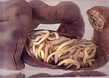
largest roundworms in the human intestine, known as nematodes.the most prevalent nematode infection in the world. A lumbricoides can grow to a length of 20 to 35 centimeters.
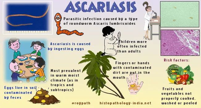
The method of transmission is via eating eggs that have been infected by human waste in the soil. After several weeks of soil incubation, eggs begin to develop into embryos.
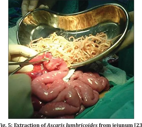
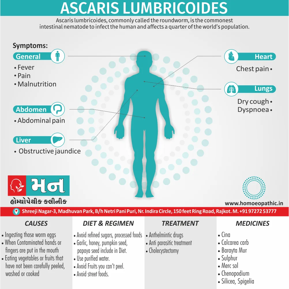
The worm load or burden affects how severe the symptoms are. One of the symptoms that arises from the larvae migrating to the lungs is acute transitory pneumonitis. Large worm burdens in the colon can cause malnutrition and bowel blockages. Adult worm migration can cause peritonitis related to intestinal perforation, duct obstructions, and appendix occlusion.
After hatching in the small intestine, larvae pass through the mucosa and are passively carried by portal blood to the liver and then the lungs. Both pneumonitis and eosinophilia are brought on by the lung migration. The larvae enter the lung through alveoli, where they are then coughed up and consumed. The mature worm stays in the digestive tract, where it deposits eggs that are expelled through the stool. Instead of attaching, they remain in their current location by moving constantly.
The worms migrate and settle in ducts when the human host becomes feverish or receives antibiotic treatment, which may restrict the flow of blood.
Usually, the diagnosis is made based on the presence of adult or egg worms in the feces. The outer surface of the oval-shaped eggs is knobby. Sputum samples taken during the lung migration can show eosinophils and larvae.
Mebendazole or pyrantel pamoate are the forms of treatment. Preventive hygiene and appropriate disposal of human waste are essential components of prevention.
Lumbricoides Ascaris Lumbricoides
largest roundworms in the human intestine, known as nematodes.the most prevalent nematode infection in the world. A lumbricoides can grow to a length of 20 to 35 centimeters.
The method of transmission is via eating eggs that have been infected by human waste in the soil. After several weeks of soil incubation, eggs begin to develop into embryos.
The worm load or burden affects how severe the symptoms are. One of the symptoms that arises from the larvae migrating to the lungs is acute transitory pneumonitis. Large worm burdens in the colon can cause malnutrition and bowel blockages. Adult worm migration can cause peritonitis related to intestinal perforation, duct obstructions, and appendix occlusion.
After hatching in the small intestine, larvae pass through the mucosa and are passively carried by portal blood to the liver and then the lungs. Both pneumonitis and eosinophilia are brought on by the lung migration. The larvae enter the lung through alveoli, where they are then coughed up and consumed. The mature worm stays in the digestive tract, where it deposits eggs that are expelled through the stool. Instead of attaching, they remain in their current location by moving constantly.
The worms migrate and settle in ducts when the human host becomes feverish or receives antibiotic treatment, which may restrict the flow of blood.
Usually, the diagnosis is made based on the presence of adult or egg worms in the feces. The outer surface of the oval-shaped eggs is knobby. Sputum samples taken during the lung migration can show eosinophils and larvae.
Mebendazole or pyrantel pamoate are the forms of treatment. Preventive hygiene and appropriate disposal of human waste are essential components of prevention.
- Published on
Differential Diagnosis of Hemolytic Autoimmune Anemia
• Nonimmune mediated: microangiopathic hemolytic anemia, hypersplenism, cardiac valve prosthesis, giant cavernous hemangiomas, march hemoglobinuria, physical agents, infections, heavy metals, drugs (nitrofurantoin, sulfonamides)
• Hemolytic anemia caused by membrane defects: paroxysmal nocturnal hemoglobinuria, spur cell anemia, Wilson’s disease
• Nonimmune mediated: microangiopathic hemolytic anemia, hypersplenism, cardiac valve prosthesis, giant cavernous hemangiomas, march hemoglobinuria, physical agents, infections, heavy metals, drugs (nitrofurantoin, sulfonamides)
• Hemolytic anemia caused by membrane defects: paroxysmal nocturnal hemoglobinuria, spur cell anemia, Wilson’s disease
- Published on
Differential Diagnosis of Drug Induced Anemia
• Drugs that may produce or promote blood loss: aspirin, alcohol, nonsteroidal anti-inflammatory agents, corticosteroids, anticoagulants
• Drugs that may interfere with red cell production by inducing marrow suppression or aplasia: alcohol, antineoplastic drugs, antithyroid drugs, antibiotics, oral hypoglycemic agents, phenylbutazone, azidothymidine (AZT)
• Drugs that interfere with vitamin B12, folate, or iron absorption or utilization: nitrous oxide, anticonvulsant drugs, antineoplastic drugs, isoniazid, cycloserine
• Drugs capable of promoting hemolysis: immune mediated, penicillins, quinine, methyldopa, procainamide, mitomycin C, oxidative stress, antimalarials, sulfonamide drugs, nalidixic acid
• Drugs that may produce or promote blood loss: aspirin, alcohol, nonsteroidal anti-inflammatory agents, corticosteroids, anticoagulants
• Drugs that may interfere with red cell production by inducing marrow suppression or aplasia: alcohol, antineoplastic drugs, antithyroid drugs, antibiotics, oral hypoglycemic agents, phenylbutazone, azidothymidine (AZT)
• Drugs that interfere with vitamin B12, folate, or iron absorption or utilization: nitrous oxide, anticonvulsant drugs, antineoplastic drugs, isoniazid, cycloserine
• Drugs capable of promoting hemolysis: immune mediated, penicillins, quinine, methyldopa, procainamide, mitomycin C, oxidative stress, antimalarials, sulfonamide drugs, nalidixic acid
- Published on
Differential Diagnosis of Hypochromic Anemia
• Thalassemia
• Other microcytic hemoglobinopathies
• Normal or increased body iron stores
• Iron deficiency anemia
• Impaired iron metabolism
• Hereditary
• Disorders of heme synthesis: sideroblastic anemias
• Disorders of globin synthesis
• Defective absorption, transport, or use of iron
• Decreased body iron stores
• Anemia of chronic disease
• Acquired
• Thalassemia
• Other microcytic hemoglobinopathies
• Normal or increased body iron stores
• Iron deficiency anemia
• Impaired iron metabolism
• Hereditary
• Disorders of heme synthesis: sideroblastic anemias
• Disorders of globin synthesis
• Defective absorption, transport, or use of iron
• Decreased body iron stores
• Anemia of chronic disease
• Acquired
- Published on
Differential Diagnosis of Iron Deficiency Anemia
• Anemia of chronic disease
• Sideroblastic anemia
• Thalassemia trait
• Lead poisoning
• Anemia of chronic disease
• Sideroblastic anemia
• Thalassemia trait
• Lead poisoning
- Published on
Differential Diagnosis of Low Reticulocyte Count Anemia
• Microcytic anemia (mean corpuscular volume [MCV] < 80): iron deficiency, thalassemia minor, sideroblastic anemia, lead poisoning
• Macrocytic anemia (MCV > 100): megaloblastic anemias, folate deficiency, vitamin B12 deficiency, drug-induced megaloblastic anemia, nonmegaloblastic macrocytosis, liver disease,
hypothyroidism
• Normocytic anemia (MCV 80–100): early iron deficiency, aplastic anemia, myelophthisic disorders, endocrinopathies, anemia of chronic disease, uremia, mixed nutritional deficiency
• Microcytic anemia (mean corpuscular volume [MCV] < 80): iron deficiency, thalassemia minor, sideroblastic anemia, lead poisoning
• Macrocytic anemia (MCV > 100): megaloblastic anemias, folate deficiency, vitamin B12 deficiency, drug-induced megaloblastic anemia, nonmegaloblastic macrocytosis, liver disease,
hypothyroidism
• Normocytic anemia (MCV 80–100): early iron deficiency, aplastic anemia, myelophthisic disorders, endocrinopathies, anemia of chronic disease, uremia, mixed nutritional deficiency
- Published on
Differential Diagnosis of Megaloblastic Anemia, due to Cobalamin Deficiency
• Usurping of luminal Cbl (inadequate Cbl binding to IF)
• Nutritional Cbl deficiency (insufficient Cbl intake): vegetarians, vegans, breast-fed infants of mothers with pernicious anemia
• Metabolic disorders (Cbl not used by cell)
• Loss or atrophy of gastric oxyntic mucosa (deficient intrinsic factor [IF] molecules): total or partial gastrectomy, pernicious anemia, caustic destruction (lye)
• Insufficiency of pancreatic protease: pancreatic insufficiency
• Inborn enzyme errors (rare)
• Inactivation of pancreatic protease: Zollinger-Ellison syndrome
• IF/post IF receptor defects: Imerslund-Graesbeck syndrome, transcobalamin (TC) II deficiency
• Drug-induced effects (Slow-K, biguanides, cholestyramine, colchicine, neomycin, p-aminosalicylic acid [PAS])
• Disorders of plasma Cbl transport (TC II–Cbl not delivered to TC II receptors)
• Disorders of ileal mucosa/IF receptors (IF-Cbl not bound to IF receptors)
• Diminished or absent IF receptors: ileal bypass, resection, or fistula
• Congenital TC II deficiency, defective binding of TC II–Cbl to TC II receptors (rare)
• By Diphyllobothrium latum
• By bacteria; stasis syndromes (blind loops, pouches of diverticulosis, strictures, fistulas, anastomoses); impaired bowel motility (scleroderma, pseudo-obstruction); hypogammaglobulinemia
• Acquired disorders: N2O inhalation
• Abnormal mucosal architecture/function: tropical or Non tropical sprue, Crohn’s disease, TB ileitis, infiltration by lymphomas, amyloidosis
• Abnormal intragastric events (inadequate proteolysis of food Cbl): atrophic gastritis, partial gastrectomy with hypochlorhydria
• Abnormal events in small bowel lumen: inadequate pancreatic protease (R-Cbl not degraded, Cbl not transferred to IF)
• Usurping of luminal Cbl (inadequate Cbl binding to IF)
• Nutritional Cbl deficiency (insufficient Cbl intake): vegetarians, vegans, breast-fed infants of mothers with pernicious anemia
• Metabolic disorders (Cbl not used by cell)
• Loss or atrophy of gastric oxyntic mucosa (deficient intrinsic factor [IF] molecules): total or partial gastrectomy, pernicious anemia, caustic destruction (lye)
• Insufficiency of pancreatic protease: pancreatic insufficiency
• Inborn enzyme errors (rare)
• Inactivation of pancreatic protease: Zollinger-Ellison syndrome
• IF/post IF receptor defects: Imerslund-Graesbeck syndrome, transcobalamin (TC) II deficiency
• Drug-induced effects (Slow-K, biguanides, cholestyramine, colchicine, neomycin, p-aminosalicylic acid [PAS])
• Disorders of plasma Cbl transport (TC II–Cbl not delivered to TC II receptors)
• Disorders of ileal mucosa/IF receptors (IF-Cbl not bound to IF receptors)
• Diminished or absent IF receptors: ileal bypass, resection, or fistula
• Congenital TC II deficiency, defective binding of TC II–Cbl to TC II receptors (rare)
• By Diphyllobothrium latum
• By bacteria; stasis syndromes (blind loops, pouches of diverticulosis, strictures, fistulas, anastomoses); impaired bowel motility (scleroderma, pseudo-obstruction); hypogammaglobulinemia
• Acquired disorders: N2O inhalation
• Abnormal mucosal architecture/function: tropical or Non tropical sprue, Crohn’s disease, TB ileitis, infiltration by lymphomas, amyloidosis
• Abnormal intragastric events (inadequate proteolysis of food Cbl): atrophic gastritis, partial gastrectomy with hypochlorhydria
• Abnormal events in small bowel lumen: inadequate pancreatic protease (R-Cbl not degraded, Cbl not transferred to IF)
- Published on
Differential Diagnosis of Megaloblastic Anemia due to Folate Deficiency
• With normal intestinal mucosa
• With mucosal abnormalities: tropical and nontropical sprue, regional enteritis
• Some drugs
• Physiologic: pregnancy and lactation, prematurity, infancy
• Pathologic: intrinsic hematologic disease (autoimmune hemolytic disease), drugs, malaria; hemoglobinopathies (Sjögren’s syndrome, thalassemia), red blood cell (RBC) membrane defects (hereditary spherocytosis, paroxysmal nocturnal hemoglobinopathy); abnormal hematopoiesis (leukemia/lymphoma, myelodysplastic syndrome, agnogenic myeloid metaplasia with myelofibrosis); infiltration with malignant disease; dermatologic (psoriasis)
• Inadequate cellular utilization: folate antagonists (methotrexate), hereditary enzyme deficiencies involving folate
• Drugs (multiple effects on folate metabolism): alcohol, sulfasalazine, triamterene, pyrimethamine, trimethoprimsulfamethoxazole, diphenylhydantoin, barbiturates
• Defective cellular folate uptake: familial aplastic anemia (rare)
• Decreased dietary intake: poverty and famine (associated with kwashiorkor, marasmus), institutionalized individuals (psychiatric/nursing homes), chronic debilitating disease/goat’s milk (low in folate), special diets (slimming), cultural/ethnic cooking techniques (food folate destroyed) or habits (folate-rich foods not consumed)
• Congenital folate malabsorption (rare)
• With normal intestinal mucosa
• With mucosal abnormalities: tropical and nontropical sprue, regional enteritis
• Some drugs
• Physiologic: pregnancy and lactation, prematurity, infancy
• Pathologic: intrinsic hematologic disease (autoimmune hemolytic disease), drugs, malaria; hemoglobinopathies (Sjögren’s syndrome, thalassemia), red blood cell (RBC) membrane defects (hereditary spherocytosis, paroxysmal nocturnal hemoglobinopathy); abnormal hematopoiesis (leukemia/lymphoma, myelodysplastic syndrome, agnogenic myeloid metaplasia with myelofibrosis); infiltration with malignant disease; dermatologic (psoriasis)
• Inadequate cellular utilization: folate antagonists (methotrexate), hereditary enzyme deficiencies involving folate
• Drugs (multiple effects on folate metabolism): alcohol, sulfasalazine, triamterene, pyrimethamine, trimethoprimsulfamethoxazole, diphenylhydantoin, barbiturates
• Defective cellular folate uptake: familial aplastic anemia (rare)
• Decreased dietary intake: poverty and famine (associated with kwashiorkor, marasmus), institutionalized individuals (psychiatric/nursing homes), chronic debilitating disease/goat’s milk (low in folate), special diets (slimming), cultural/ethnic cooking techniques (food folate destroyed) or habits (folate-rich foods not consumed)
• Congenital folate malabsorption (rare)
